Download
1 / 48
480 likes | 720 Views
15.1 Origin of Life. There are three possibilities for the appearance of the first living organisms on Earth 1. Extraterrestrial origin Life was transferred to Earth from a distant planet 2. Special creation Life was created by supernatural or divine forces 3. Evolution

E N D
15.1 Origin of Life • There are three possibilities for the appearance of the first living organisms on Earth • 1. Extraterrestrial origin • Life was transferred to Earth from a distant planet • 2. Special creation • Life was created by supernatural or divine forces • 3. Evolution • Life may have evolved from inanimate matter, with selection as the driving force • Only the third possibility is scientifically testable
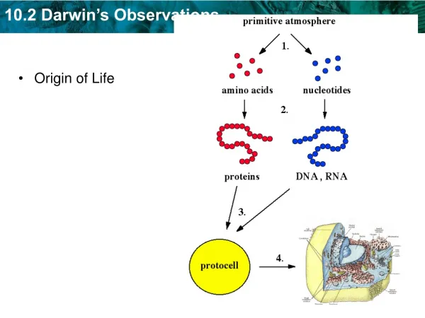
Fig. 15.1 Forming Life’s Building Blocks • Life originated 2.5 billion years ago • The Earth’s atmosphere then had no oxygen • It was rich with hydrogen-rich gases (NH3, CH4) • These simple molecules combined to form more complex molecules • Lightning provided the energy
Forming Life’s Building Blocks • Stanley Miller and Harold Urey reconstructed the oxygen-free atmosphere of early Earth in their lab • Cellular building blocks form spontaneously, when the system is subjected to lightning or UV light • They concluded that life may have evolved in a “primordial soup” of biological molecules
Forming Life’s Building Blocks • Concerns have been raised about the “primordial soup” hypothesis • No oxygen => no protective ozone layer • Therefore, the UV light would have destroyed the essential ammonia and methane gases • Louis Lerman, in 1986, proposed the bubble model • Key chemical life-building processes took place within bubbles on the ocean’s surface • Inside the bubbles the essential gases would be protected from UV light
15.2 How Cells Arose • The first step may have been the formation of tiny bubbles termed microspheres • These have cell-like properties • Those microspheres better able to incorporate molecules and energy persisted longer than others • The first macromolecule produced was RNA • It provided a possible early mechanism of inheritance • Later, RNA was replaced as hereditary material by the much more stable double-stranded DNA
Earth is formed 4.5 billion years ago Fig. 15.3 A clock of biological time
15.3 The Simplest Organisms • The fossil record indicates that prokaryotes appeared 2.5 billion years ago • Eukaryotes, on the other hand, appeared only 1.5 billion years ago • Today prokaryotes are the simplest and most abundant form of life on earth • Prokaryotes occupy a very important place in the web of life in earth
Spiral Rod Fig. 4.9 Spherical The Structure of a Prokaryote • Prokaryotes are small, simply organized, single cells that lack a nucleus • Prokaryotes include the bacteria and archaebacteria • They come in three main shapes • Rod-shaped (bacilli) • Spherical (cocci) • Spirally coiled (spirilli)
The Structure of a Prokaryote • The cell membrane of prokaryotes is encased in a cell wall • Bacterialcell walls are composed ofpeptidoglycan • Network of polysaccharides linked together by peptide cross-links • Archaebacterialcell walls lack peptidoglycan • They are made of proteins, sugars or both
Gram-positive • Have a thick peptidoglycan layer • Lack outer membrane • Gram-negative • Have a thin peptidoglycan layer • Have an outer membrane containing lipopolysaccharide • Bacteria can be divided into two groups based on their cell wall architecture The name refers to a differential stain developed by Hans Christian Gram • Gram-positive cells retain the primary crystal violet stain • Gram-negative cells don’t and are stained by a safranin (red) counterstain
Many bacteria have capsules • A gelatinous layer found external to the cell wall • Many bacteria possess threadlike flagella • Long external appendages used for locomotion • Some bacteria also possess pili • Short external appendages used for attachment • In harsh conditions, a few bacteria can form endospores • Highly-resistant structures that may germinate into active bacteria when conditions improve
Fig. 7.1 • Prokaryotes reproduce by binary fission
Fig. 15.5 Both cells contain a complete copy of the plasmid • Some bacteria undergo a process called conjugation • The undirectional transfer of plasmid DNA following cell-to-cell contact Pilus connecting the two cells together
Prokaryotes differ from eukaryotes in many respects They have very little internal organization They are unicellular and much smaller They possess a single chromosome They are far more metabolically diverse 15.4 Comparing Prokaryotes to Eukaryotes
Prokaryotic Metabolism • Prokaryotes have evolved many more ways than eukaryotes to acquire carbon and energy • Acquisition of Carbon • Autotrophs = Use CO2 as their only carbon source • Heterotrophs = Use preformed organic compounds as carbon sources • Acquisition of Energy • Phototrophs = Use light as energy source • Chemotrophs = Use chemicals as energy source
Prokaryotic Metabolism • Based on carbon and energy sources, prokaryotes can be divided into four categories • 1. Photoautotrophs • Use the energy of sunlight to build organic molecules from CO2 • Cyanobacteria • 2. Chemoautotrophs • Obtain energy by oxidizing inorganic substances • Nitrifiers oxidize ammonia or nitrite
Prokaryotic Metabolism • Based on carbon and energy sources, prokaryotes can be divided into four categories • 3. Photoheterotrophs • Use light as energy and pre-formed organic molecules as carbon sources • Purple nonsulfur bacteria • 4. Chemoheterotrophs • Use organic molecules as carbon and energy sources Decomposers and most pathogens
15.5 Importance of Prokaryotes • Prokaryotes were largely responsible for creating the properties of the atmosphere and soil found on Earth • They are the principal decomposers • They are the only organisms capable of fixing nitrogen, thus making it available for others • They cause major diseases in plants and animals, including humans
Fig. 15.7 Exxon Valdez oil spill • Prokaryotes are instrumental in genetic engineering • Nonpolluting insect control agents • Removing environmental pollutants
15.6 Prokaryotic Lifestyles • Prokaryotes belong to two domains • Archaea • Members are termed archaebacteria • Bacteria • Members are termed bacteria • Includes the vast majority of prokaryotes described
Hot spring in Yellowstone National Park Fig. 15.8 15.6 Prokaryotic Lifestyles • Archaeabacteria include • Methanogens • Use H2 to reduce CO2 to CH4 • Live in swamps and marshes • Extremophiles • Live in unusually harsh environments • Thermoacidophiles • Hot and acidic environments
Fig. 15.9 15.6 Prokaryotic Lifestyles • Bacteria include • Cyanobacteria • Individual cells adhere in filaments • The most prominent photosynthetic bacteria • Also capable of nitrogen fixation • In specialized cells called heterocysts Heterocysts
15.6 Prokaryotic Lifestyles • There are numerous phyla of non-photosynthetic bacteria • Some are chemoautotrophs • But most are heterotrophs • Decomposers • Bacteria cause many diseases in humans • Among the most serious is tuberculosis (TB) • It is caused by Mycobacterium tuberculosis • The emergence of drug-resistant strains in the 1990s has raised serious medical concerns
15.7 The Structure of Viruses • Viruses are not living organisms • Rather, they are “parasitic” chemicals that can only reproduce within living cells • Viruses are very small • Range from 17 – 1,000 nm • Viruses occur in all organisms • In every case, the basic structure is the same • Segments of DNA or RNA wrapped in a protein coat called the capsid • There is considerable difference, however, in the details
Membrane-like layer Fig. 15.10 The structure of bacterial, plant and animal viruses Not found in all viruses Capsid
Bacteriophages are viruses that infect bacteria The most studied are double-stranded DNA bacteriophages, including Phage T4 Fig. 15.11b 15.8 How Bacteriophages Enter Prokaryotic Cells Tail fibers • Phage lambda (l) • Same basic structure as T4 • But only possesses one thin tail fiber
Bacteriophages undergo two types of reproductive cycles Lytic cycle Example: T4 bacteriophage Lysogenic cycle Example: Bacteriophage l 15.8 How Bacteriophages Enter Prokaryotic Cells
Lytic Cycle • Bacteriophage uses tail fibers to attach to bacterial cell wall • Tail contracts and DNA is injected into host cytoplasm • Viruses multiply within infected cell • They eventually lyse (rupture) the cell and spread to other cells • Viruses that can only undergo a lytic cycle are termed virulent
Uninfected cell Lysis of cell Bacterial chromosome Assembly of new viruses using bacterial cell machinery Virus attaching to cell wall Lytic cycle Replication of virus Viral DNA injected into cell Fig. 15.12 Lytic and lysogenic cycles of a bacteriophage
Lysogenic Cycle • Some bacteriophages do not immediately kill the cells they infect • Rather, they integrate their DNA into the chromosome of their host cell • This phenomenon is termed lysogeny • The viral DNA is called the prophage • At a later time, the prophage may exit the genome • This initiates viral replication and, ultimately, cell lysis • Viruses that can become stably integrated within their host’s genome are termed temperate
Uninfected cell Lysis of cell Bacterial chromosome Assembly of new viruses using bacterial cell machinery Virus attaching to cell wall Lytic cycle Replication of virus Viral DNA injected into cell Reduction to prophage Prophage exits the bacterial chromosome Lysogenic cycle Reproduction of lysogenic bacteria Fig. 15.12 Lytic and lysogenic cycles of a bacteriophage Viral DNA integrated into bacterial chromosome
Lysogenic Conversion • Expression of prophage genes may have an important effect on the host cell • The bacterium Vibrio cholerae exists in two forms • A harmless form • A virulent form that causes the disease cholera • This form is infected with a temperate bacteriophage which carries a gene encoding the cholera toxin • Lysogenic conversion is also responsible for the presence of toxin genes in other bacteria • Corynebacterium diphtheriae Diphtheria
Plant viruses enter plant cells through tiny rips in their cell wall at points of injury Animal viruses enter their host cells through endocytosis A diverse array of viruses occur among animals We will focus on the human immunodeficiency virus (HIV) 15.9 How Animal VirusesEnter Cells
Fig. 15.13 HIV • HIV causes Acquired Immunodeficiency Syndrome (AIDS) • AIDS was first reported in the US in 1981 • Clinical symptoms of AIDS until generally 8-10 years after infection with HIV • During this latent period, carriers of HIV are infectious
HIV • Attachment and entry • Virus circulates throughout the entire body but will only infect macrophages • Involved in the uptake and recycling of organic material • Specificity is mediated by gp120 protein spikes found on the HIV surface • gp120 binds to CD4 marker on the macrophages • It then binds a second receptor protein, called CCR5 • This triggers endocytosis of the virus into the macrophage
HIV • Replication • Once inside macrophages, HIV sheds its protective coat, releasing its RNA genome • The viral enzyme reverse transcriptase synthesizes double-stranded DNA from the single-stranded RNA • The enzyme does not proofread so many mutations are introduced • The double-stranded DNA is integrated into the host cell’s genome • In all of this, no lasting damage is done to the host cell
HIV • Starting AIDS • Eventually, by chance, HIV alters the gene for gp120 causing it to produce a new form of gp120 protein • This prefers to bind to a different co-receptor, called CXCR4 • The virus can now infect CD4+ T cells • New viruses exit T cells by bursting through the plasma membrane • The destruction of the body’s T cells causes the onset of AIDS • Cancers and opportunistic infections are free to invade the defenseless body
15.10 Disease Viruses • Influenza • Perhaps the most lethal virus in human history • Natural reservoirs ducks and pigs in central Asia • AIDS (HIV) • First entered humans from chimpanzees in Africa • The chimpanzee virus is called simian immunodeficiency virus (SIV) • Ebola virus • Filamentous virus that attacks connective tissue • Natural host of the virus is unknown
15.10 Disease Viruses • Hantavirus • Discovered in 1993 • Natural host is the deer mice • SARS • Severe acute respiratory syndrome • Caused by a coronavirus • Natural host is most likely the civet • West Nile Virus • Mosquito-borne virus • Natural host is birds
Fig. 15.15 Thelast known smallpox case 15.10 Disease Viruses Ali Maow Maalin of Somalia • Smallpox • Caused by the variola virus • 15 million worldwide cases in 1967 • A world immunization campaign eradicated the disease in 1977 • The virus still exists in Atlanta and Moscow • “Weaponized” smallpox remains a bioterrorist threat