Download
1 / 21
210 likes | 308 Views
From the Human Eye Down to Microscopes at the Nano-Scale. Michael Crocker Valerie Goss Pat Mooney Rebecca Quardokus. Outline. Discussion of Optics History Ranges of Visual Resolution Scanning Electron Microscopy (SEM) Fundamentals Operation Scheme Atomic Force Microscopy (AFM)

E N D
From the Human Eye Down to Microscopes at the Nano-Scale Michael Crocker Valerie Goss Pat Mooney Rebecca Quardokus
Outline Discussion of Optics History Ranges of Visual Resolution Scanning Electron Microscopy (SEM) Fundamentals Operation Scheme Atomic Force Microscopy (AFM) Scanning Tunneling Microscopy (STM)
Scanning Bunny A distant cousin of the energizer bunny who helps to explain nanoscience concepts. WELCOME PENN HIGH SCHOOL
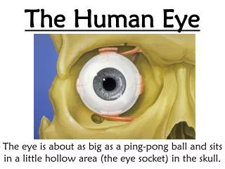
How do we see an object? source target detector …and often you’ll need a lens
Requirements of Vision • The light that reaches the eye must have a color between red (760nm) and blue (400nm) – or a mixture of these colors • The light that reaches the eye must be sufficiently bright – usually requires a sufficiently bright source www.uvabcs.com/uvlight-typical.php , August 31, 2009 UV UV UV C B A visible light infrared 760 wavelength in nm 3000 290 320 760 400
Seeing Atomic Structure • Light must be about 0.1nm in wavelength to see atomic structure: x-rays • But our eyes can’t detect x-rays - 0.1nm light - (5000 times smaller wavelength than we can see) • Options • Use x-rays and detector (to replace the eye) • Use particles (e.g. electrons) and detector • Electrons of the appropriate wavelength are easier to produce and direct than light – Scanning Electron Microscope (SEM) • Alternate imaging techniques • Atomic Force Microscope (AFM) • Scanning Tunneling Microscope (STM)
Scanning Electron Microscope Michael Crocker
Let’s bounce something else at the surface! e- e- e- e- e- e- e- e- e- e- e- Basic Idea? Animal sight and traditional microscopes collect deflected light Some are “reflected” Some are absorbed
Electron Beam Column Beam created from heated filament Beam travels through a vacuum Electro-magnetic fields act as lenses Electron beam hits the sample in a precise location Scattered and “secondary” electrons are detected Beam scans back and forth http://bioweb.usu.edu/emlab/TEM-SEM%20Teaching/How%20SEM%20works.html
Electrons Hit Surface and Detection Primary electrons come from the beam Some scatter back, others dislodge electrons http://www4.nau.edu/microanalysis/Microprobe-SEM/Signals.html
Example Images http://gsc.nrcan.gc.ca/labs/ebeam/sem_gallery_e.php
Atomic Force Microscope Valerie Goss
What is the AFM? An analogue! We can sense with our hands by touching.
AFM cantilever and AFM tips www.veeco.com
The powerful, versatile AFM Resolutions: X and Y 2 -10 nm Z 0.05 nm Microstructure of solids: CD, glass beads, circuits Biological samples: skin cross section, viruses, bacteria, blood, DNA and RNA ~30 um scan www.nanotech-now.com/.../antonio-siber.htm Aug 27, 2009
Scanning Tunneling Microscope Rebecca Quardokus
Electrons tunnel! With a higher probability than cars STM measures the current created by tunneling electrons Scanning Tunneling Microscopy (STM) Images courtesy of http://www.ieap.uni-kiel.de and www.renault.com
Scanning Tunneling Microscopy (STM) C60 “Bucky Balls” Each C60 diameter is ~ 10Å 1 Å = 1x 10⁻¹⁰ m Image courtesy of http://nano.tm.agilent.com
Scanning Tunneling Microscopy (STM) Xenon on Nickel Individual atoms? That’s small! Iron on Copper Images courtesy of http://www.almaden.ibm.com