Download
1 / 5
50 likes | 51 Views
Researchers analyze biofilm samples rich in zinc sulfide to understand the relationship between organic material, metals, and bioremediation potential. They discover proteins in the biofilm, suggesting a key role in aggregation and extracellular biomineralization.

E N D
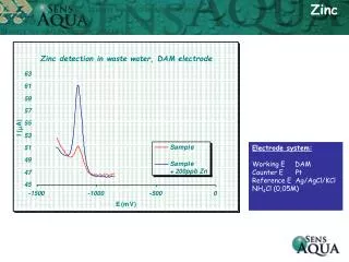
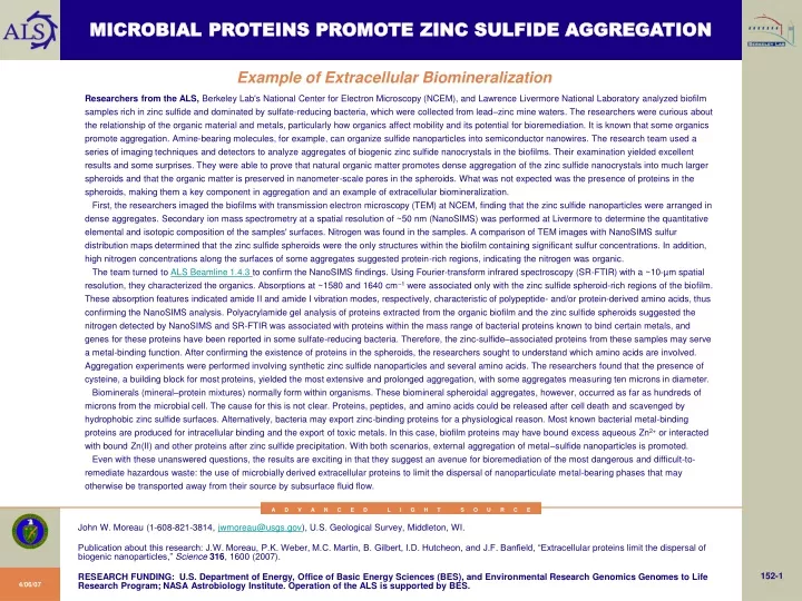
MICROBIAL PROTEINS PROMOTE ZINC SULFIDE AGGREGATION Example of Extracellular Biomineralization Researchers from the ALS, Berkeley Lab's National Center for Electron Microscopy (NCEM), and Lawrence Livermore National Laboratory analyzed biofilm samples rich in zinc sulfide and dominated by sulfate-reducing bacteria, which were collected from lead–zinc mine waters. The researchers were curious about the relationship of the organic material and metals, particularly how organics affect mobility and its potential for bioremediation. It is known that some organics promote aggregation. Amine-bearing molecules, for example, can organize sulfide nanoparticles into semiconductor nanowires. The research team used a series of imaging techniques and detectors to analyze aggregates of biogenic zinc sulfide nanocrystals in the biofilms. Their examination yielded excellent results and some surprises. They were able to prove that natural organic matter promotes dense aggregation of the zinc sulfide nanocrystals into much larger spheroids and that the organic matter is preserved in nanometer-scale pores in the spheroids. What was not expected was the presence of proteins in the spheroids, making them a key component in aggregation and an example of extracellular biomineralization. First, the researchers imaged the biofilms with transmission electron microscopy (TEM) at NCEM, finding that the zinc sulfide nanoparticles were arranged in dense aggregates. Secondary ion mass spectrometry at a spatial resolution of ~50 nm (NanoSIMS) was performed at Livermore to determine the quantitative elemental and isotopic composition of the samples' surfaces. Nitrogen was found in the samples. A comparison of TEM images with NanoSIMS sulfur distribution maps determined that the zinc sulfide spheroids were the only structures within the biofilm containing significant sulfur concentrations. In addition, high nitrogen concentrations along the surfaces of some aggregates suggested protein-rich regions, indicating the nitrogen was organic. The team turned to ALS Beamline 1.4.3 to confirm the NanoSIMS findings. Using Fourier-transform infrared spectroscopy (SR-FTIR) with a ~10-µm spatial resolution, they characterized the organics. Absorptions at ~1580 and 1640 cm–1 were associated only with the zinc sulfide spheroid-rich regions of the biofilm. These absorption features indicated amide II and amide I vibration modes, respectively, characteristic of polypeptide- and/or protein-derived amino acids, thus confirming the NanoSIMS analysis. Polyacrylamide gel analysis of proteins extracted from the organic biofilm and the zinc sulfide spheroids suggested the nitrogen detected by NanoSIMS and SR-FTIR was associated with proteins within the mass range of bacterial proteins known to bind certain metals, and genes for these proteins have been reported in some sulfate-reducing bacteria. Therefore, the zinc-sulfide–associated proteins from these samples may serve a metal-binding function. After confirming the existence of proteins in the spheroids, the researchers sought to understand which amino acids are involved. Aggregation experiments were performed involving synthetic zinc sulfide nanoparticles and several amino acids. The researchers found that the presence of cysteine, a building block for most proteins, yielded the most extensive and prolonged aggregation, with some aggregates measuring ten microns in diameter. Biominerals (mineral–protein mixtures) normally form within organisms. These biomineral spheroidal aggregates, however, occurred as far as hundreds of microns from the microbial cell. The cause for this is not clear. Proteins, peptides, and amino acids could be released after cell death and scavenged by hydrophobic zinc sulfide surfaces. Alternatively, bacteria may export zinc-binding proteins for a physiological reason. Most known bacterial metal-binding proteins are produced for intracellular binding and the export of toxic metals. In this case, biofilm proteins may have bound excess aqueous Zn2+ or interacted with bound Zn(II) and other proteins after zinc sulfide precipitation. With both scenarios, external aggregation of metal–sulfide nanoparticles is promoted. Even with these unanswered questions, the results are exciting in that they suggest an avenue for bioremediation of the most dangerous and difficult-to-remediate hazardous waste: the use of microbially derived extracellular proteins to limit the dispersal of nanoparticulate metal-bearing phases that may otherwise be transported away from their source by subsurface fluid flow. A D V A N C E D L I G H T S O U R C E John W. Moreau (1-608-821-3814, jwmoreau@usgs.gov), U.S. Geological Survey, Middleton, WI. Publication about this research: J.W. Moreau, P.K. Weber, M.C. Martin, B. Gilbert, I.D. Hutcheon, and J.F. Banfield, “Extracellular proteins limit the dispersal of biogenic nanoparticles,” Science316, 1600 (2007). 152-1 RESEARCH FUNDING: U.S. Department of Energy, Office of Basic Energy Sciences (BES), and Environmental Research Genomics Genomes to Life Research Program; NASA Astrobiology Institute. Operation of the ALS is supported by BES. 4/06/07
MICROBIAL PROTEINS PROMOTE ZINC SULFIDE AGGREGATION Example of Extracellular Biomineralization • ZnS nanocrystals in biofilms of sulfate-reducing bacteria • Aggregate into larger spheroids • Containing proteins • Evidence of extracellular biomineralization • SR-FTIR analysis at ALS Beamline 1.4.3 • Amide I (~1640 cm–1 ) and II (~1580 cm–1) absorption features • Diagnostic of amino-acid–associated bond vibrations in polypeptides and/or proteins • Associated only with ZnS spheroid-rich regions • Confirming NanoSims detection of organic N • And determining existence of proteins • Experiments show cysteine has most extensive and prolonged aggregation • Exciting news for bioremediation • Use of microbially derived extracellular proteins • To limit mobility of metallic waste A D V A N C E D L I G H T S O U R C E J.W. Moreau (University of California, Berkeley), P.K. Weber and I.D. Hutcheon (Lawrence Livermore National Laboratory), M.C. Martin and B. Gilbert (Berkeley Lab), and J. Banfield (University of California, Berkeley, and Berkeley Lab).[Science 316, 1600 (2007)] 152-2
MICROBIAL PROTEINS PROMOTE ZINC SULFIDE AGGREGATION Example of Extracellular Biomineralization TEM (top) and NanoSIMS (bottom) images of biogenic zinc sulfide aggregates. Red, green, and blue areas represent regions of sulfur, nitrogen, and carbon, respectively. Orange and yellow areas show the intimate association of both sulfur and nitrogen. A D V A N C E D L I G H T S O U R C E J.W. Moreau (University of California, Berkeley), P.K. Weber and I.D. Hutcheon (Lawrence Livermore National Laboratory), M.C. Martin and B. Gilbert (Berkeley Lab), and J. Banfield (University of California, Berkeley, and Berkeley Lab).[Science 316, 1600 (2007)] 152-3
MICROBIAL PROTEINS PROMOTE ZINC SULFIDE AGGREGATION Example of Extracellular Biomineralization NanoSIMS secondary-ion images of an ultramicrotomed TEM section of biofilm. Nitro-gen was detected as CN–, NO–, and NS–, and was quantified by comparison to reference samples. Composite element distribution map (~10 µm x 10 µm) of 12C (blue), 12C14N for N (green), and 32S (red). Colors reflect the proportion of each species. Uniformly red regions represent relatively pure S (as ZnS), whereas orange and yellow regions indicate the presence of increased levels of N. Light blue regions indicate the presence of both C and N, with little to no S (no ZnS). A D V A N C E D L I G H T S O U R C E J.W. Moreau (University of California, Berkeley), P.K. Weber and I.D. Hutcheon (Lawrence Livermore National Laboratory), M.C. Martin and B. Gilbert (Berkeley Lab), and J. Banfield (University of California, Berkeley, and Berkeley Lab).[Science 316, 1600 (2007)] 152-4
MICROBIAL PROTEINS PROMOTE ZINC SULFIDE AGGREGATION Example of Extracellular Biomineralization SR-FTIR transmission spectra of biogenic ZnS aggregates (black) and background biofilm (gray). Amide I (~1640 cm–1) and II (~1580 cm–1) absorption features are diagnostic of amino-acid–associated bond vibrations in polypeptides and/or proteins. A D V A N C E D L I G H T S O U R C E J.W. Moreau (University of California, Berkeley), P.K. Weber and I.D. Hutcheon (Lawrence Livermore National Laboratory), M.C. Martin and B. Gilbert (Berkeley Lab), and J. Banfield (University of California, Berkeley, and Berkeley Lab).[Science 316, 1600 (2007)] 152-5