Download
1 / 25
281 likes | 538 Views
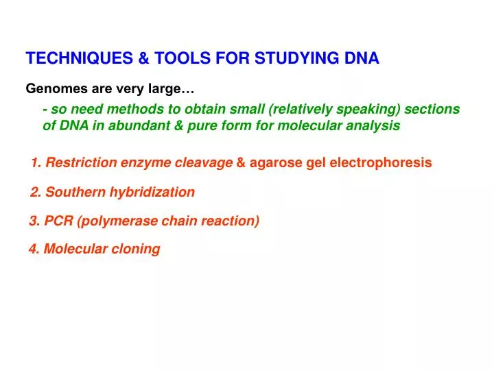
TECHNIQUES & TOOLS FOR STUDYING DNA. Genomes are very large…. - so need methods to obtain small (relatively speaking) sections of DNA in abundant & pure form for molecular analysis. 1. Restriction enzyme cleavage & agarose gel electrophoresis. 2. Southern hybridization.

E N D
TECHNIQUES & TOOLS FOR STUDYING DNA Genomes are very large… - so need methods to obtain small (relatively speaking) sections of DNA in abundant & pure form for molecular analysis 1. Restriction enzyme cleavage& agarose gel electrophoresis 2. Southern hybridization 3. PCR (polymerase chain reaction) 4. Molecular cloning
1. Restriction enzymes - cleave DNA into specific, small fragments 1. DNA endonucleases cut double-stranded DNA at specific recognition sites (often palindromic) “blunt” ends “sticky” ends Fig.2.10 BamHI: staggered cut with 5’ overhang 2. Recognition sequences often 4 or 6 bp, but also “rare cutters” (eg. NotI 5’ GCGGCCGC 3’) can be useful for generating very large fragments in genomic mapping Sites are often shown as one strand, but implicit that double-stranded
3. Two different restriction enzymes may generate same “sticky ends” • “compatible ends” useful for cloning • (eg. partial Sau3A genomic digest ligated into BamHI site in vector) 4. Isoschizomers – restriction enzymes with identical recognition sequences … but may have different response to methylation state MspI cleaves 5’ CCGG 3’ regardless of methylation state HpaII does not cleave 5’ CCGG 3’ if 2d C is methylated
Example using isoschizomers to assess DNA methylation state of genes in cancer patients - used assay called “HpaII tiny fragment Enrichment by Ligation–mediated PCR” - found “significant aberrancy in promoter methylation patterns compared with normal NBCs” NBC: naïve B cells (ie. from healthy people) log(HpaII/MspI) ratios “Genome-wide DNA methylation analysis reveals novel targets for drug development in mantle cell lymphoma” Leshchenko et al. Blood 116:1025, 2010
Agarose gel electrophoresis - to separate DNA fragments by size Fig.T2.2 Small fragments migrate more rapidly than large ones Fig.T2.1
“Pulsed field” electrophoresis for separation of large DNA molecules • For example: • - restriction fragments generated by “rare cutters” • megaplasmids • whole chromosomes (eg. yeast) Fig.3.30
But if DNA is very complex, number of fragments (of various sizes) generated is too large to see discrete bands after electrophoresis... so “continuum” of signals in lane 1 2 3 Why are different profiles expected for genomic DNA cleaved with BamHI (6 bp cutter) vs. Sau3A (4 bp cutter)? B S Distance (on average) expected between restriction sites depends on probability of occurrence of that sequence 50 kb - 20 kb - 10 kb - 5 kb - Enzyme with 6 bp recognition site expected to cut a DNA molecule (of 50% GC content) on average once every 46 bp (ie. 4096 bp) (see p.86) 2 kb - 1 kb - 0.5 kb - Lane 1 = uncut DNA Lane 2 = 6 bp cutter Lane 3 = 4 bp cutter
2. Southern hybridization - to detect specific restriction fragment containing sequence (eg.gene) of interest(vs. all other fragments) Fig.2.11A • After electrophoresis, denature DNA and transfer it from gel to membrane (eg. by capillary action or electroblotting…) …so that DNA fragments remain in same relative positions
- probe will anneal with single-stranded DNA on blot, if sequences are complementary Fig.2.11B 2. Hybridize blot with “probe” (DNA, oligomer, cDNA, or RNA… which is tagged either radioactively or non-radiolabelled) and detect specific hybrid by autoradiography Stability of hybrid depends on: - length of hybrid, (eg for oligomer probes), GC content … - hybridization conditions (such as temperature, ionic strength…)
Some applications of Southern blot analysis - to identify restriction fragment carrying sequence (eg gene) of interest - to identify gene copy number (eg. multi-gene families) Aside: Northern hybridization – RNA is electrophoresed, blotted to membrane and hybridized with probe (eg gene of interest) - to determine if gene is active, size of mRNA & its abundance … (Fig.5.11) (to be discussed in Topic 6)
3. PCR - polymerase chain reaction - to obtain one specific DNA region in large copy number - rapid amplification of DNA region of interest by enzymatic reaction in test tube 1.Denaturation of duplex DNA 2. Annealing of 2 different primers (synthetic oligomers, usually 15-25 nt ) • flank region of interest, • in opposite orientation … so anneal to opposite strands of DNA 3. Extensionof complementary strands - cycle repeated 25-30 times First cycle Video at http://www.maxanim.com/genetics/PCR/pcr.swf http://www.youtube.com/watch?v=x5yPkxCLads Fig.2.28 "Scientists for Better PCR" a Bio-Rad Music Video for the all new 1000-Series Thermal Cyclers”
Subsequent PCR cycles - discrete PCR product generated - its length corresponds to distance between primers (including the primers) - sequences at ends of amplicon correspond to the 2 primers used Fig.2.29
Designing PCR primers: 5’ 3’ 3’ 5’ …GATTCC... 5’ …GCGTAT... 3’ 5’ …CTAAGG… …CGCATA... 3’ Typically use 20-25 nt oligomers, but for simplicity (as on a test) 6’mers are shown here Why choose ~ 20’mers? Tip: see Question 2.5 in text (p.61) - for specificity… How many times would a particular 20’mer sequence be expected to be present in the human genome, by chance? Why choose ~ 50% GC? (and avoid homopolymeric stretches) - to reduce chance of non-specific annealing at other genomic sites
How to double-check that PCR product (amplicon) is correct one? 1. Is it the right size? - agarose gel electrophoresis (with size markers) Well Fig. 2.30 2. Does it contain the right sequence (eg gene X)? - Southern hybridization - using gene X (eg. clone) as probe - restriction analysis - are expected restriction sites present? - nested PCR New primer - design “internal” primers to use in 2d PCR experiment with 1st PCR product as template DNA New primer
3’ 5’ 3’ 5’ RT-PCR 3’ 5’ 3’ 5’ 3’ 5’ Gene-specific oligomer or oligo dT 5’ 3’ 5’ 3’ 5’ 3’ - then sequence RT- PCR product directly (or after cloning) 5’ 3’ - (need sequence data to design primers for RT-PCR) 3’ 5’ (see p.142, Chapter 5)
Real-time quantitative RT-PCR - detection and measurement of products generated during each cycle of PCR by using a reporter fluorescent probe eg. SYBR green, TaqMan - to measure relative or absolute amount of mRNA present in different tissue types/developmental stages/environmental conditions… ΔRn : increment of fluorescent signal at each time point CT : PCR cycle number where reporter fluorescence is greater than threshold RFU = relative fluorescence units NTC = no template control http://www.ncbi.nlm.nih.gov/projects/genome/probe/doc/TechQPCR.shtml NCBI Technologies website
Powers & pitfalls of PCR - rapid method to generate large amounts of specific segment of DNA (product usually < 10 kb in length) - need very small amount of template DNA …but can lead to contamination problems - need prior sequence info to design primers Some applications of PCR: - genomic analysis - RNA studies (RT-PCR) - forensic work - paleobiology (“ancient” DNA)
4. CLONING - to obtain one specific DNA region in large copy number - by using host cell (eg. E.coli) to amplify DNA of interest - DNA fragments ligated intovector … then introduced into bacterial (or yeast…) cell by transformation to generate clone library = collection of clones whose inserts cover the entire genome Aside: cDNA library - mRNAs reverse-transcribed into cDNAs and cloned (Fig. 5.32) Fig. 2.15
Examples of cloning vectors used to generate clone library (or bank) Table 2.4 • Plasmid • - to clone < 10 kb fragments • - origin of replication, selectable markers eg. antibiotic resistance in bacteria: ampicillin, tetracycline … or nutrient requirement in yeast: URA3, TRP1 … Insert disrupts lacZ’ gene, so Xgal on plate not converted to blue colour & colonies are white lacZ’ = marker for rapid screening of recombinants Fig.2.18
2. Phage lambda - to clone 15-20 kb DNA fragments (Aside: also l vectors for cloning cDNAs of 1-5 kb) Mid-region of l DNA molecule can be removed and replaced with similar-sized insert DNA of interest, then packaged in phage particle 3. Cosmid - l-plasmid hybrid, cos site to package DNA in phage particle - to clone ~40-45 kb fragments 4. BAC - bacterial artificial chromosome (~8 kb) with F (fertility) plasmid origin of replication - to clone ~ 300 kb fragments - most commonly used vector for cloning large DNA fragments
5. YAC - yeast artificial chromosome - to clone ~ 1 Mbp fragments - but sometimes DNA rearrangements & instability of inserts Fig.2.25 Selectable markers (TRP1 & URAS3) - yeast host strain requires tryptophan & uracil in medium to grow, but transformants (which possess TRP1 & URA3 genes on YAC) can grow in medium lacking them
How many clones needed in library to cover a complete genome? Depends on: genome size and insert size in vector Number of clones N that must be screened to isolate a given sequence with a probability of P: N = ln (1 – P) ) ln (1 – insert length/ genome length) Table 2.4 Rule-of-thumb: For 99% probability of success, the total # bp present in clones screened must be about 5 x greater than total genome size For E.coli (genome size ~ 4.6 Mbp), how many clones needed if average insert size = 10 kb?
Strategies to generate overlapping clones? - use in assembling genomic maps 1. Use two clone banks with restriction fragments derived from differentrestriction enzymes (or from incomplete digestion with one restriction enzyme) A ABAB BABA A BA B … … DNA cleaved with B DNA cleaved with A A A A A B B B B A A etc. B B A A B B etc. Can use clone from B library as probe to find clone in A library that contains part of the same sequence… prepare library B B prepare library A A A A B B plus neighbouring sequences… A A B B Then can look for overlapping clone in B library... “chromosome walking”
2. Random fragmentation of DNA (eg sonication or nebulization ) then blunt-end ligation into vector (or ligation into vector after linkers containing restriction sites added) 1 kb Recover DNA of desired size (eg. 1 kb) by gel electrophoresis (or repeated nebulization) & prepare clone library ... having random overlapping segments of genome Nebulizer for random shearing of DNA Cold Spring Harbor Protocols 2010 Aside: this method was used to obtain the first complete bacterial genome sequence (Haemophilus influenza) (Topic 5 & Fig.4.10)