Download
1 / 35
360 likes | 836 Views
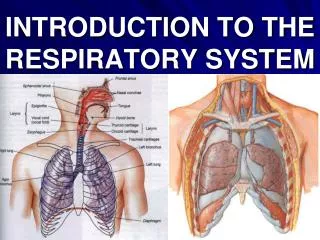
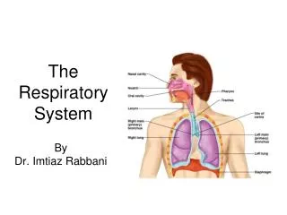
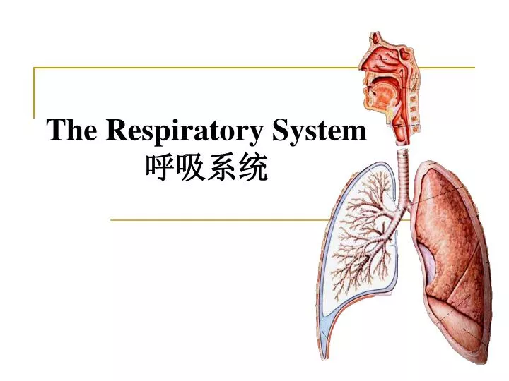
The Respiratory System 呼吸系统. 目的和要求 掌握 鼻腔的分部及各部的形态结构。 掌握 鼻旁窦的位置及开口部位。 掌握 喉的位置、喉软骨的名称 掌握 喉口的围成、喉腔的分部和各部的形态结构。 掌握 左、右主气管的形态差别。 掌握 肺的位置、形态和分叶。 掌握 肺根的构成及各结构的排列关系。 掌握 胸膜下界与肺下界的体表投影;胸膜顶的体表投影。 了解纵隔的概念及分部。. The General Description. The respiratory system includes the res-

E N D
目的和要求 • 掌握鼻腔的分部及各部的形态结构。 • 掌握鼻旁窦的位置及开口部位。 • 掌握喉的位置、喉软骨的名称 • 掌握喉口的围成、喉腔的分部和各部的形态结构。 • 掌握左、右主气管的形态差别。 • 掌握肺的位置、形态和分叶。掌握肺根的构成及各结构的排列关系。 • 掌握胸膜下界与肺下界的体表投影;胸膜顶的体表投影。 • 了解纵隔的概念及分部。
The General Description The respiratory system includes the res- piratory tract and lungs.The primary function of this system is to supply the body with oxygen and get rid of excess carbon dioxide resulting from cell metabolism. Nose Pharynx upper respiratory Larynx tract 上呼吸道 Trachea lower respiratory tract Bronchi 下呼吸道
Section 1 The Nose 鼻 I.External nose: Root of nose Back of nose Apex of nose Ala of nose II.Nasal cavity –is divided into two halves by the nasal septum. Little region
Two parts: Nasal vestibule鼻前庭 Nasal cavity proper 固有鼻腔 Lateral wall Nasal conchae鼻甲 Nasal meatus鼻道 Sphenoethmoidal recess 蝶筛隐窝
Remove the middle nasal conchae Semilunar hiatus 半月裂孔 Ethmoidal infundibulum 筛漏斗 Ethmoidal bulb 筛泡 Nasolacrimal canal 鼻泪管
Mucous membrane of nose • Olfactory region嗅区 • Respiratory region呼吸区
Frontal sinus Ethmoidal sinuses Sphenoid sinus Maxillary sinus III.The paranasal sinuses and their site of drainage into the nose
咽 • 1、概述:上宽下窄,前后略扁的漏斗状肌性器官。上起颅底,下达第六颈椎下缘,全长12cm,是消化道、呼吸道的共同通道。咽的前壁不完整,分别与鼻腔、口腔、喉腔相通。 • 2、分部:软腭下缘、会厌上缘将咽分为鼻咽、口咽、喉咽三部分。
1)鼻咽:顶的后部有咽扁桃体,婴幼儿发达,6~7岁后萎缩,10岁退化。在咽的侧壁距下鼻甲约1cm处有一三角形的裂孔为咽鼓管咽口。其前上后隆起为咽圆枕,它与咽后壁之间为咽隐窝,是鼻咽癌的好发部位,其底为破裂孔,可向颅内扩散。1)鼻咽:顶的后部有咽扁桃体,婴幼儿发达,6~7岁后萎缩,10岁退化。在咽的侧壁距下鼻甲约1cm处有一三角形的裂孔为咽鼓管咽口。其前上后隆起为咽圆枕,它与咽后壁之间为咽隐窝,是鼻咽癌的好发部位,其底为破裂孔,可向颅内扩散。 • 2)口咽:侧壁有腭扁桃体窝,内有腭扁桃体。 • 咽淋巴环:咽扁桃体、腭扁桃体、舌扁桃体在鼻腔、口腔通咽处所形成的环状结构,具有防御功能。 • 3)喉咽:前方有喉口,两侧有梨状隐窝,为异物停留处。
Section 2 The Larynx The larynx is a part of respiratory passage as well as the orgen of phonation. Position:It lies in the anterior part of the neck (below the hyoid bone), and extends from vertebral level of C3 to C6.
I.Laryngeal cartilages (I)Thyroid cartilage 甲状软骨Shield-shaped Laryngeal prominence (II)Arytenoid杓状软骨 Paired, pyramid shaped (III)Cricoid cartilage 环状软骨 Complete ring shaped like a signet ring
(IV)Epiglottic cartilage 会厌软骨 leaf-shaped elastic cartilage
II.Laryngeal joints 1.Thyrohyoid membrane 甲状舌骨膜 2.cricothyroid joint 环甲关节 3.cricoarytenoid joint 环杓关节
4.Quadrangular membrane 方形膜 5.Conus elasticus弹性圆锥 • vocal ligament声韧带 • Median cricothyroid ligament环甲正中韧带 6.Cricotracheal ligament 环状软骨气管韧带
III.Muscles of larynx 喉肌IV .Laryngeal cavity 喉腔 (I)Aditus laryngis 喉口 bounded by upper border epiglottis; aryepiglottic folds and interarytenoid notch
Structure features 1.Two folds : Vestibular folds 前庭襞 Vocal folds 声襞 2.Two fissures Rima vestibuli 前庭裂 Fissure of glottis 声门裂 Intermembranous partIntercartilagrnous part ..
(II)Threeparts: Laryngeal vestibule 喉前庭 Intermedial cavity of larynx 喉中间腔 Infraglottic cavity 声门下腔
Section 3 The Trachea and Bronchi I.Trachea气管It is the passage for air. It extends from the lower border of the circoid cartilage at the level of sixth cervical vertebra to the level of the sternal angle. Structure features • Consists of about 14-17 C-shaped incomplete tracheal cartilages • Carina of trachea 气管隆嵴
II.Bronchi Right principal bronchus 右主支气管 Shorter, wider, and morevertical Left principal bronchus 左主支气管 Narrower, longer, and more horizontal
Section 4 The Lungs The lungs are situated one on each side within the thorax,and separated from each other by the heart and other contents of the mediastinum. The External features: Cone-shaped An Apex of lung A Base of lung Three surfaces Three borders
left Medial surface 1.Hilum of lung 肺门 2.Root of lung 肺根 • Order of structures: From before backward: R.L.- V.A.B. From above downward: • R.-B. A. V. • L.-A. B. V. right
Lobes and Fissure 1.Right lung • Two fissures : horizontal and oblique • Three lobes 2.Left lung • One fissure : oblique • Two lobes
III. The Bronchial tree支气管树IV .Bronchopulmonary segments支气管肺段
Section 5The PleuraThe pleura is thin,glistening,slippery serous membrane that lines the inner surface of thorax and the surfaces of lungs. General features: • Two layers Visceral pleura - adheres to lung Parietal pleura- lines the thoracic cavity
pleural cavity: Two pleural layers continuing with each other at root of lung formed a closed potential space
I.Named parts of parietal pleura Cupula of pleura Costal pleura Mediastinal pleura Diaphragmatic pleura
II.Pleura recesses • Costodiaphragmatic recesse 肋隔隐窝 • Costomediastinal recess 肋纵隔隐窝 • Phrennicomediastinal recess 隔纵隔隐窝
III.The surface projection of inferior margins of lung and pleurae
Section 6The mediastinum 1.Definition 2.Divisions
Summary and question: 1.临床上常把哪几部分称为上呼吸道? 2.鼻旁窦有哪些?分别开口于何处? 3.喉的软骨有哪些?喉腔可分为哪几部分? 4.为什么气管异物多坠入右侧? 5.肺根内有哪些结构出入,其位置关系如何? 6.壁胸膜可分为哪几部分?胸膜发生炎症时,渗出液常易积聚于何处?