Download

1 / 46
890 likes | 1.78k Views
Tumor Markers. Overview. Cancer remains the second leading cause of death in the USA, behind heart disease, with an estimated 1,437,180 new cases & 565,650 deaths in 2008 alone. It is expected that malignancies of prostate & breast origin will be

E N D
Overview Cancer remains the second leading cause of death in the USA, behind heart disease, with an estimated 1,437,180 new cases & 565,650 deaths in 2008 alone. It is expected that malignancies of prostate & breast origin will be the most common causes of new diagnoses in men & women, respectively, with tumors of the lung & bronchus the leading cause of cancer-related death in both genders
Cancer A simple definition of cancer is: A relatively autonomous growth of tissue, that can develop into a solid mass or tumor & spread to other areas of the body Cancer cells : • Are not subject to regulatory system of cell growth • Infiltrate adjacent tissue (in contrast to benign tumours) • Form metastases due to lymphogenic or haematogenic spread The proliferation of normal cells is thought to be regulated by growth promoting proto-oncogenes & counterbalanced by tumor-suppressor genes.
Cell cycle Phases of cell activity divided into G, S & M (Growth, DNA synthesis & Mitosis respectively) Apoptosis Programmed cell death Development of new blood vessels to supply oxygen & nutrients to cells
Proto-oncogenes • Proto-oncogenes are genes present in normal human cells & their products (proteins) may play important roles in normal cellular processes (as normal promotion of growth) • These proto-oncogenes may be activated to oncogenes • Oncogenes can convert normal cells into tumor cancer cells. • In cancer cells, oncogenes may be: Identical to normal genes but regulation of their expression in cancer cells is abnormal Or: Show very small structural differences from their counterparts in normal cells. These differences will produce agents that leads to transformation of normal cells to cancer cells.
Tumour Suppressor Genes • Tumour suppressor genes are genes that encode for proteins (product) that are involved in protecting cells from unregulated growth • Mutation in tumor suppressor genes may lead to cancer
How to Diagnose Cancer? • A golden dream of an oncologist is to diagnose cancer at an early stage. • Diagnosis of carcinoma in the early stages is a difficult task due to: - Lack of symptomsat early stages. - Diagnostic procedures (X ray, CT, MRI..) are not suitable for early diagnosis. Methods only detect tumors of at least 1-2 cm in size (1x10⁹cells) or more. • Tumor markers are considered as one of the highly valuable tools for detection & follow up of malignant tumors & their secondaries.
Tumor Markers Tumor Markers Substances present in or produced by a tumor itself OR Produced by host in response to a tumor Can be used to Determine the presence of a tumor ORDifferentiate a tumor from normal tissue Based on Measurements in blood or secretions
Tumor Markers Tumor markers include a variety of substances like: • Oncofetal proteins • Cell surface antigens • Cytoplasmic proteins • Hormones • Enzymes • Receptors • Oncogenes & their products (proteins)
Characteristics of an Ideal Tumor Marker • Should be highly specific (negative in all negative cases)…. Diagnosis & Screening • Should be highly sensitive (positive in all positive cases)….Diagnosis & Screening • Should provide a lead-time over clinical diagnosis….Screening • The levels of the marker should correlate with the tumor burden .. Prognosis • It is accurately reflecting any tumor progression or regression….Monitoring • Procedure of estimation of the marker should be reliable • The test used for detection should be relatively cheap
Applications of Tumor Markers 1- Diagnosis(D): to help to establish the diagnosis 2- Screening(S): to identifypatients with early cancer (before appearance of clinical manifestations) 3- Prognosis(P): to assess the aggressiveness 4- Monitoring (M): follow up of regression or progression Evaluate the Response to Treatment (RT) Detection of Recurrence (R) 5- Determination of Risk: i.e. in genetically predisposed individ.
1- Screening Screening asymptomatic individuals (in very early cases of cancer) Use of tumor markers, to date, has generally not been an effective strategy due to: • Most of the clinically used tumor markers are found in normal cells & benign conditions in addition to cancer cells. • The relatively low prevalence of individual cancer types. • The screening role is generally very useful if restricted to groups with risk factors as: - Family history - Environmental exposure - Geographic prevalence - Clinical profile
1- Screening cont. High risk groups: are individuals at increased risks for malignancy Tumor markers used for screening of high risk individuals: • Alpha fetoprotein (AFP) For patients with liver cirrhosis or chronic hepatitis who are at risk for development of hepatocelluar carcinoma • Prostate-specific antigen (PSA) For men older than 50 years in conjunction with a physical examination (clinical profile) for early detection of cancer prostate • Calcitonin For first degree relatives of a patient (family history) with medullary carcinoma
1- Screening cont. Sensitivity & Specificity of Screening Tumor Markers: Ideal tumor marker for screening asymptomatic population should be: 100% sensitive: Always positive in patients with the disease 100% specific: Always negative in individuals who do not have the disease For examples: If a test gives positive results in 99 patients out of 100 patients: Its sensitivity is 99% If a test gives negative results in 90 normal individuals out of 100 normal individ. Its specificity is 90%
1- Screening cont. In reality, an ideal tumor marker which gives 100% specificity & 100 % sensitivity does notexist. To increase sensitivity & specificity of a tumor marker • Combination of multiple tumor markers • Combination of tumor markers with other procedures e.g. combination of Carbohydrate Antigen 125 (CA 125) with ultrasonography for early detection of ovarian malignancy
2- Prognosis • For cancer patients, determination of prognosis is based on determination of aggressiveness of tumor, which , in turn determines how a patient should be treated (surgery, chemotherapy, radiotherapy, etc) • As the serum concentrations of tumor markers increases with progression and usually reaches the highest levels when tumors become metastasized, the serum levels at diagnosis are likely to reflect the aggressiveness of the tumor and predict the outcome. • High levels of serum tumor marker measured during diagnosis would indicate the presence of a malignant or metastatic tumor associated with poor prognosis.
4- Monitoring of Disease, Response of Treatment & Detection of Recurrence • Constitutes the most common clinical use of serum tumor markers. • Markers usually increase with progressive disease, decrease with remission & do not change significantly with stable disease. • After treatment (surgical resection, radiation or chemotherapy), tumor markers are routinely followed serially (to monitor the response to treatment & recurrence). • It is desirable to monitor the patient using a highly sensitive tumor marker test to detect recurrence as early as possible • The appearance of most of the circulating tumor markers have a lead time of several month (3-6 months) prior to the stage at which many of the physical procedures can be used for detection of the cancer • So, rising tumor marker levels may detect recurrence of disease before any clinical or radiological evidence of disease is apparent (biochemical recurrence)
5- Determination of Risk • Usually involves genetic probes that evaluate any specific genetic abnormality or mutation noted to indicate an increased risk of a particular malignancy. • Examples of such abnormalities would include: - Carriers of Philadelphia chromosome for hematological malignancies - Carriers of BRCA 1 or 2 genes, which confer a higher risk of breast or ovarian malignancies
Factors that affect serum concentrations of tumor markers • False positive results occur with: • Inflammatory conditions • Benign conditions • Presence of liver diseases: Causes disturbances in metabolism and excretion of some tumor markers • Disturbances of renal function: affects levels of some tumor markers • Consequences of diagnostic and therapeutic procedures: digito-rectal examination, mamography, surgery, radio and chemotherapy • As a consequence of different physiological conditions: as in pregnancy may affect ßHCG, AFP…..).
AFP: alpha fetoprotein B-hCG: beta human chorionc gonadotrpin CEA: carcinembrionic antigen CA 125: carbohydrate antigen 125
Factors that affect serum concentrations of tumor markers False negative results occur with: • Insufficient expression of the marker or production of the marker in some of the tumor cells only • Insufficient blood circulation in the tumor • Production of autoantibodies against the marker • Rapid degradation & clearance of the marker
Frequency of Ordering of Markers • As a general guideline, the time interval between serial determinations should be 3 months. • But in case of an abnormal value, a repeat estimate can be ordered within 2 to 4 weeks irrespective of the initial reading. Checking the effectiveness (success ) of treatment: • Confirming of the success of surgical removal of a tumor is checked by measuring the marker concentrations ideally after a period not less than 5-6 half-lives(to allow tumor marker levels to fall to normal). • In case of treatment with chemotherapy or radiotherapythe period should be longer
GUIDELINES FOR ORDERING/ INTERPRETING TUMOR MARKER TESTS • Never rely on the result of a single test • Order every test from the same laboratory • Consider half-life of the tumor when interpreting the result • Consider how the Tumor Marker is removed or metabolized
Examples of Frequently Ordered Tumor Markers • Alpha-fetoprotein • CA-125 • CEA • hCG • PSA • Her-2/neu • p53 • BrCa1 • BrAa2 • CA-15.3 • CS-19.9 • Estrogen & progesterone receptor • VMA
Oncofetal Proteins • These are proteins produced during fetal life. • They are present in high concentrations in the sera of fetuses & decrease to low levels or disappear after birth. • They reappear in individuals with cancer. • This demonstrates that certain genes are reactivated as a result of the malignant transformation of cells. • There are several oncofetal antigens as AFP & CEA
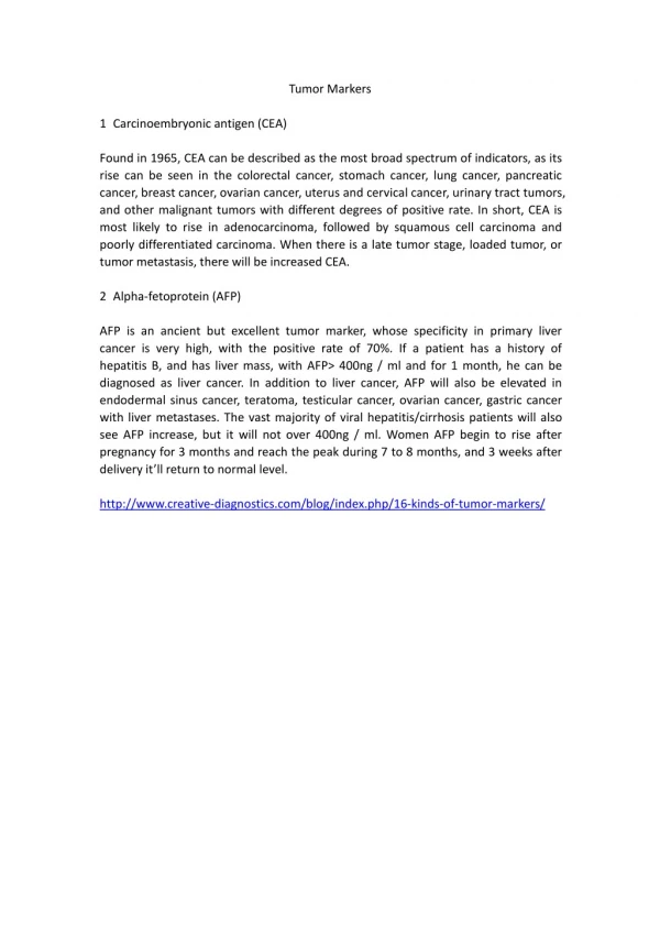
Alfa-feto Protein (AFP) • AFP is a glycoprotein. • It is one of the major proteins in the fetal circulation • The fetal AFP reaches the peak at 14 weeks of gestation & declines at term. • The maternal AFP level increase from 12 weeks to peak during the third trimester • It is a marker of hepatocellular carcinoma & nonseminomatous carcinoma of testis • Other causes of increased AFP due to non-malignant causes: - Pregnancy - Non- cancerous liver diseases : hepatitis & cirrhosis • Except in pregnancy , AFP level greater than 1000ug/L indicates cancer
Alfa-fetoprotein (AFP) cont. • Clinical Application of AFP: • Screening, diagnosis, prognosis & monitoring of hepatocellular carcinoma (i.e. hepatoma). • Used with ultrasound imaging every 6 months in patients at high risk of developing HCC e.g. patients with hepatitis B virus & hepatitis C virus-induced liver cirrhosis AFP is used for early detection (in the lead period) which is ~ 3 - 6 months before clinical manifestations of the cancer appear. • AFP is not completely specific for HCC • AFP may be increased in pregnancy & benign liver disease. • AFP is used as a tumor marker for nonseminomatous testicular cancer (in combination with -hCG)
Carcinoembryonic Antigen (CEA) It is the most widely used tumor marker for colorectal cancer. The main clinical use of CEA is as a tumor marker for colorectalcancer for: Prognosis Post-surgery surveillance Monitorresponse to chemotherapy
Hormones as Tumor marker The production of hormone in cancer involves 2 separate routes: The endocrine tissue that normally produces the hormone can produce excessive amounts (e.g. Vinylmandilic acid, VMA is a tumor marer for pheochrmocytoma, tumor of the adrenal medulla) 2. A hormone can be produced by nonendocrine tissue that normally does not produce the hormone (e.g. ectopic production of ACTH & PTH)
Human Chorionic Gonadotropin (hCG) • hCG is a hormone normally secreted by trophoblasts in the placenta during pregnancy. It is a glycoprotein consisting of and subunits. • It is the most useful marker for detection of gestational trophoblastic diseases(GTDs) that include: • Hydatiform mole (vesicular mole) • Choriocarcinoma • It is alsoelevated in nonseminomatous tumors of the testis (with AFP).
Enzymes as Tumor Markers Generally, an increase in enzymes is not specific or sensitive to identify type of cancer. PSA is an exception
Prostate Specific Antigen (PSA) • PSA is a glycoprotein produced only in the epithelial cells of the acini & ducts of the prostate. There are 2 major forms of PSA that are found in the blood: Free & Complexed: to 1-antichymotrypsin or 2-macroglobulin. • Total PSA isused in screening & in monitoring of prostate cancer • Free PSA can help to differentiate levels of PSA that are in the grey zone: i.e. not high enough to diagnose cancer prostate, but not low enough to rule out the diagnosis of cancer prostate: Patient with cancer prostate have a lower % of free PSA.
Prostate Specific Antigen (PSA) • Annual PSA testing for screening of prostate cancer is indicated for: • In men over 50 years old • In younger men at high risk: e.g. with a family history of prostate cancer. • To increase the accuracy of the PSA testing, it is essential to use age-adjusted cutoff values of PSA • The best clinical use & first clinical applications of PSA testing was to monitor for the progression of prostate cancer after therapy (e.g. radical prostatectomy) • Causes other than prostate cancer that can elevated PSA: • Prostate infection • Prostate irritation • Benign prostatic hyperplasia (enlargement)
Carbohydrate Markers Carbohydrate relate tumor markers: are either: Antigens on tumor cells Or: Secreted by the tumor cells ( CA = carbohydrate antigens)
Carbohydrate Antigen-125 (CA-125) CA-125 may be useful for detecting ovarian tumors: Only clinically accepted serologic marker of ovarian cancer Diagnoseovarian tumors at early stages Distinguish benign masses from ovarian cancer Monitoring treatments CA-125 is notconsidered specific enough for ovarian cancer, as it may be elevated in patients with Endometriosis During the first trimester of pregnancy During menstruation.
Estrogen/Progesterone Receptors (ER/PR) • These receptors can play a role in breast carcinogenesis assessment of ER/PR status is crucial for optimal treatment planning (similar to HER-2) • Decreased recurrence rates & disease-related mortality have been demonstrated with tamoxifenfor ER+ cancers, which are not seen when the tumors are ER– • Receptor status also has significant prognostic implications, with best (ER+/PR+ ) to poorest (ER−/PR–.)
Genetic Markers Two classes are involved in the development of cancer Oncogenes: as HER-2/neu Suppressor genes: as p53, BRCA1 , BRCA2…
Human Epidermal Growth Factor Receptor 2 (Her-2/neu) • HER-2 gene productis a transmembrane protein normally involved in cell growth & differentiation through its interaction with circulating growth factors • It is identified as an proto-oncogeneas its amplification results in protein over expression that supports rapid cellular proliferation (i.e. becomes oncogene). • Her-2/neu is a marker for breast & ovarian cancers • In breast cancer to determine the type of therapy: • Breast cancer positive for Her-2/neu is responsive to treatment (Herceptin) • Breast cancer negative for Her-2/neu is NOT responsive to treatment
Tumor suppressor genes Tumour suppressor genes are genes that encode for proteins that are: • Involved in normal protection of cells from unregulated • growth • If mutated may lead to cancer • Examples of tumor suppressor genes: • P53 • BRCA1 & BRCA2
p53 • p53 (an example of tumor suppressor genes) P53 gene is located on ch.17 (together with genes of BRCA1 & Her-2/neu) Gene product (protein) normally result in cell cycle arrest & induces apoptosis • Upon mutation: loss of function mutation cancer: Cancer colon, Brain cancer, Cancer lung & Cancer liver Carcinogens causing mutations in p53: • AflatoxinB Potent hepatocarcinogen (common in China & Africa) Causes mutations of p53 gene leading to cancer liver • Smoking: Causes mutations in p53 gene leading to lung cancer
BRCA1 & BRCA2 • They are genes that are inherited as autosomal dominant trait. • Two genetic loci : BRCA1 on chromosome 17 & BRAC2 on chromosome 13 • If these genes are mutated, cancer occur (mutation is inherited to offspring) • Carriers of BRCA1 gene mutation: have an 85% risk of developing breast cancer & 45% risk of developing ovarian cancer