Download
1 / 50
520 likes | 947 Views
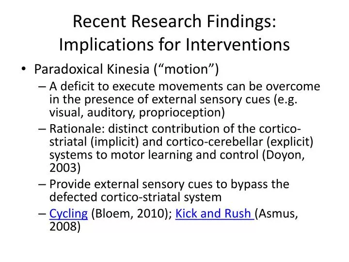
Recent Research Findings: Implications for Interventions. Paradoxical Kinesia (“motion”) A deficit to execute movements can be overcome in the presence of external sensory cues (e.g. visual, auditory, proprioception)

E N D
Recent Research Findings:Implications for Interventions • Paradoxical Kinesia (“motion”) • A deficit to execute movements can be overcome in the presence of external sensory cues (e.g. visual, auditory, proprioception) • Rationale: distinct contribution of the cortico-striatal (implicit) and cortico-cerebellar (explicit) systems to motor learning and control (Doyon, 2003) • Provide external sensory cues to bypass the defected cortico-striatal system • Cycling (Bloem, 2010); Kick and Rush (Asmus, 2008)
PTP 512Neuroscience in Physical TherapySensation and Perception Min H. Huang, PT, PhD, NCS Reading Assignments S & W: 131-132, 134-135 L-E: 108, 109, 113, 114, 136-141, 286-293
Objectives • Compare somatosensory receptor neurons relative to their size and myelination • Describe the sensory function controlled by neurons in each of the sensory pathways • Sketch the sensory homunculus • Describe the role of posterior parietal cortex (areas 5 & 7) in sensory perception • Apply knowledge of the sensory and perceptual pathways to determine expected sensory loss or impairment with injury to the peripheral nerve, spinal cord, brainstem, or cortex
Receptive Field for Cutaneous Sensation • Receptive field is the area that innervated by a single afferent neuron. • Where you may find the smallest receptive field in the body?
End Discuss at your table group • Identify the regions with impaired sensation following injury to the C8 root of brachial plexus vs. ulnar nerve distal to the wrist vs. medial brachial cutaneous nerve distal at the shoulder. • Are the affected regions the same or different and why? • Lundy’s Fig 6-4
End Discuss at your table group • What is dermatome? • Compare and contrast the regions innervated by the dermatomes vs. cutaneous nerves. Why are they different? • Lundy’s Figure 6-5
End Discussion at your table group • Compare and contrast somatosensory receptor neurons relative to their axon size and myelination • Describe the sensory function controlled by neurons in each of the sensory pathways Lundy’s Table 6-1
End Produced by Dave Foord
Large Diameter Neurons • Largediameter axons conduct faster than small diameteraxons • less resistance to electrical current in larger axons • Largediameter axons are myelinated • allows “saltatory” conduction of action potentials • Large diameter axons are affected before small diameteraxons in nerve compression injuries like carpal tunnel syndrome
End Discussion at your table group Describe the sensory function controlled by neurons in each of the sensory pathways
Sensory Pathways • Discriminative touch • Where does it cross? • Lundy’s CD: Touch • Fast pain • Where does it cross? Is it the same for the fast pain pathways innervating the trunk vs. face? • Lundy’s CD: Fast pain
Types of Peripheral Nerve Injuries Commonly Seen in Clinical Practice • Stretch injuries • Most common • PN are inherently elastic (20% stretch is OK, will not injure nerve) • e.g. Brachial plexus birth-related injuries • Laceration/severance • e.g. injury caused by a knife blade http://orthoinfo.aaos.org/topic.cfm?topic=a00077
Types of Peripheral Nerve Injuries Commonly Seen in Clinical Practice • Compression injuries • Due to mechanical compression and ischemia • Larger myelinated nerve fibers are more susceptible than smaller unmyelinated nerve fibers. • e.g. carpal tunnel syndrome, Saturday night palsy (entrapment of the nerve)
Compression Injuries: Changes in Sensory Function • Large myelinated fibers are affected first with initial sparing of smaller pain, thermal, and ANS fibers • Sensory loss typically occurs in this sequence • conscious proprioception / discriminate touch • cold • fast pain • heat • slow pain • When compression is relieved, paresthesia often develops
Compression Injuries: Changes in Motor Function • Difficult to identify the lesion in non-severe cases because of extensive intermingling of muscle fibers of different motor units in compression injuries • Innervated muscles become fatigue faster than normal • A patient with partial denervation lesion may be able to recruit enough motor units to generate a single max voluntary contraction to be graded as "normal" on MMT • Need to test for muscle fatigue
Compression Injuries: Changes in ANS Function • ANS symptoms are rare. Usually single nerve is involved unless the nerve is completely severed • Symptoms include: • lack of sweating • loss of sympathetic control of smooth muscle fibers leading to edema • if multiple nerves are involved, may have difficulty regulating BP, HR, sweating, bowel & bladder function
PNS Structure http://missinglink.ucsf.edu/lm/IDS_101_histo_resource/images/141Cx1_copy.jpg
Pathology of PNS Injury: Neurapraxia (Class I) • Temporary impairment of a local nerve conduction. Mild mechanical disruption of a nerve (e.g. carpal tunnel syndrome) • Very subtle and localized demyelination without loss of axonal continuity may be present, typically without Wallerian degeneration http://harvester.lib.utah.edu/
Pathology of PNS Injury: Neuroapraxia (Class I) • Larger fibers are affected (e.g. efferent fibers to αMN). • Small sensory fibers are not affected • Clinical features • Marked reduction in muscle strength in a specific distribution distal to the lesion • Sensation may be similarly affected • Nerve conduction across the lesion site is slowed or absent, whereas above and below the lesion it is normal • Recovery in hours to weeks
Pathology of PNS Injury: Axonotmesis (Class II) • Caused by severe compression or stretching that results in a loss of axonal continuity but endoneurial, perineurium, and epineurium are intact • Leading to Wallerian degeneration (i.e. axons and myelin degenerate) distal to the site of injury within 48 hours of a severe injury http://harvester.lib.utah.edu/
Pathology of PNS Injury: Axonotmesis (Class II) • Axonal regeneration may occur at a rate of about 1 mm/day • Clinical features • Muscle weakness or paralysis • Up to 50% atrophy in 2 weeks • Sensory loss in a specific distribution • Conduction velocities across lesion site are lost immediately • Distal to the lesion are lost within a few days
Wallerian Degeneration Normal Injury Anterograde degeneration Loss of connection with muscle Total degeneration Axons re-sprout using remaining Schwann cells as guides and re-innervate the muscle. http://www.medscape.com/viewarticle/480071_4
Two alternative models explaining the greater axon degeneration distal to the lesion (center). ‘Dying back' model (top): axonal degeneration starts at the distal end. Focal lesion model (bottom): focal lesions can trigger Wallerian degeneration of distal axons. http://www.nature.com/nrn/journal/v6/n11/fig_tab/nrn1788_F1.html
Pathology of PNS Injury: Neurotmesis (Class III) • A nerve injury characterized by loss of axonal and all connective tissue continuity • Usually results from a rapid stretch/avulsion injury or complete severance of the nerve • Clinical features • Similar to axontomesis but more severe. The prognosis is poor • Surgical repair is necessary for any chance of recovery
Patterns of somatosensory impairments • Peripheral nerve: nerve distribution • Nerve root: dermatome • Spinal cord: may involve different sensory tracts, loss of sensation usually begins one or two segments below the level of the lesion because of branching of the afferent fibers in the spinal cord • Brainstem: may have mixed ipsilateral/contralateral sensory loss of the body and the face • Cortical and subcortical structure above midbrain: contralateral body/face • Thalamus: contralateral body/face • Somatosensory cortex: contralateral body/face
Sensation vs. Perception Sensation Perception The process of integrating, organizing and interpreting sensations. Creating meaningful patterns from sensory information Require attention, memory, integration of stimuli, motivation, expectations etc. • “Picking up” sensory information • The process of detecting a stimulus (e.g. light waves-vision, sound waves-heating, chemical molecules-taste, heat or pressure-touch).
Thalamus “inner chamber” or “bedroom” in Greek“egg-shaped” grey matter structureThe last synaptic site before sensory information reaches cerebral cortex
Thalamic Connections • In addition to processing sensory information, thalamus is a major sensory relay station • Each sensory modality (except for olfactory), including vision, hearing, taste, and somatic sensation, has a different nuclear area in the thalamus, where synapses occur before the information is relayed to the cerebral cortex.
Thalamic Connections • Reciprocal projections between thalamus and cortex • Non-sensory pathways also relay their information in the thalamus before reaching the cerebral cortex, including motor inputs from basal ganglia and cerebellum, inputs from limbic system and brainstem reticular formation. • What is the clinical implications of this?
Sensory Disturbances Associated with Thalamic Lesions • Lesions of the thalamus result in decreased or lost sensation from the contralateral body or face. • Thalamic pain syndrome (central post stroke pain syndrome) • People with stroke often develop “thalamic pain syndrome” • Burning, pricking, aching, lacerating, shooting pain • Pain can be spontaneous or evoked by mechanical or thermal stimuli (e.g. touching a sheet or cold surface)
Primary Sensory Cortex Lundy’s textbook, 2007
Primary Sensory Cortex: Function Lundy’s textbook, 2007
Sensory Association Cortex Lundy’s textbook, 2007
Sensory Association Cortex: Function Unimodal sensation (one type of sensory modality) Higher-order (cortical) sensation Lundy’s textbook, 2007
Posterior Parietal Cortex (Areas 5 & 7) • Cortical sensation • Area 5 integrates information from different body parts • Area 7 also received processed visual information • Area 7 combines eye-limb information for visually guided movements (e.g. reach to grasp)
Primary Sensation Cortical or Higher-Order Sensation Graphesthesia Stereognosis Tactile extinction • Pain • Temperature • Vibration • Joint position sense • Two point discrimination • Intact primary sensation with deficits in cortical sensation such as agraphesthesia or astereognosis suggests a lesion in the ______ • Note, however, that severe cortical lesions can cause deficits in primary sensation as well.
Graphesthesia is the ability of the patient to identify characters that are written on the skin using a dull pointed object. Stereognosis is the ability to identify objects that are placed in the hand when the eyes are closed
Tactile extinction (Double Simultaneous Stimulation) • Touching homologous parts of the body on one side, the other side or both sides at once with the patient identifying which side or if both sides are touched with their eyes closed. • If the patient neglects one side on extinction, it indicates dysfunction of the contralateral posterior parietal lobe http://library.med.utah.edu/neurologicexam/html/sensory_normal.html
Complex Perceptual Dysfunction: Visual-Spatial Deficits • Spatial relations disorders encompass a constellation of impairments that have in common a difficulty in perceiving the relationship between the self and two or more objects. • Research suggests that the right parietal lobe plays a primary role in space perception. • Thus, a visual-spatial deficits most commonly occurs in patients with right-sided brain lesions with left hemiparesis.
Perceptual Dysfunction: (a)somatognosia • Body schema is a postural model of the body, including the relationship of body parts to each other and the relationship of the body to the environment • (a)somatognosia is the failure to recognize one’s parts and their relationship to each other. Attitudes towards one’s body can be indifference, delusion, or critical • e.g. hemispatial neglect http://neuroexam.com/neuroexam/content.php?p=10
Hemispatial Neglect Not absolute but a gradient of neglect Eye movements during visual search in an individual with left-sided neglect attempting to find letter Ts among Ls. Red dots-visual fixations. Yellow lines- saccadic eye movements. http://www.scholarpedia.org/article/Hemineglect
Hemispatial Neglect • Most common in damage to right posterior parietal cortex or frontal cortex From: Neuroscience: Exploring the Brain by Bear, Connors, Paradeso
Apraxia • Most common in lesion of the left frontal and posterior parietal cortex • Ideomotor apraxia • Inability to carry out an action in response to verbal command, in the absence of any comprehension deficit, motor weakness, or incoordination • Patients can make certain gestures/movements spontaneously but have trouble in making these same gestures/movements if asked to do so http://neuroexam.com/neuroexam/content.php?p=9
Apraxia • Ideational apraxia • Inability to carry out an action, either automatically or on command • Patients are unable to describe verbally how tooth-brushing is done and unable to brush the teeth either on command or automatically