Download

1 / 16
230 likes | 534 Views
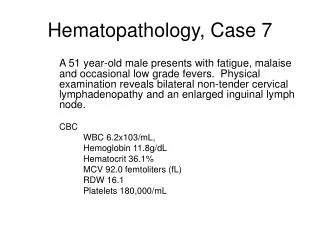
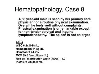
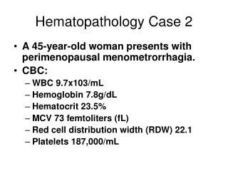
Hematopathology. Peripheral Blood Film Examination. Normal RBC : The normal human erythrocytes are biconcave disc, 7.2 Mm in diameter, and the thickness of 2.4 Mm at the periphery and 1 Mm in the center.

E N D
Peripheral Blood Film Examination • Normal RBC : The normal human erythrocytes are biconcave disc, 7.2 Mm in diameter, and the thickness of 2.4 Mm at the periphery and 1 Mm in the center. • The biconcave shape render the red cell quite flexible so that they can pass through capillaries whose minimum diameter is 3.5 Mm • Normal red cells (normochromic): have uniformly coloured haemoglobin in side the cell with a small clear paler region in the center
Shape variationAcanthocyteswith irregular, thorny speculated membrane surface projections bulbous round endsCause: abetalipoproteinemia, renal failure, liver disease, haemolytic anaemia
Ecchinocytes: cells with 10-30 uniformly distributed spiculesCause: blood loss (acute), burns, DIC, carcinoma of stomach
Elliptocytes: have a cigar shapeCause: hereditary elliptocytosis, leukemia, thalassaemia
Sickle cells:cells have a sickle with appoint at one end Cause: sickle cell anaemia, haemoglobin S disease
Spherocytic cells:are circular ( not biconcave), smaller , more condensed than normal Cause: hereditary spheroytosis, autoimmune hemolytic anaemia, septicemia
Stomatocytes:cells are cup shaped with an abnormal area of central pallor that may be oval, elongated, or slit likeCause: liver disease, alcoholism.
Target cells:cells have an increased ratio of surface to volumeCause: iron deficiency, liver disease, haemoglopinopathies, post splenectomy
Tear drop poikilocyte: cells have teardrop or pear shape Cause: myelofibrosis, extramedullary haemopoiesis, myeloid metaplasia
Size variation: • Normal: normal size ( 80- 95 ), and normal diameter (6-8u) called Normocytes • Macrocytes: increase size of cells having diameter > 8 u and MCV > 95u • Microcytes : decrease size of cells having diameter < 6 u and MCV < 80u
Content Of Structure Variation Basophilic stippling: appearanceof fine blue dots scattered in red cellsCause: haemoglopinopathies, lead poisoning, haemolytic anaemia, myelodysplasia
Heinz bodies: are denaturedparticles of haemoglobin attached to RBC membrane that appear when stained with cresyl blueCause: G6PD deficiency, drug induced
Howell jolly body:are nuclear fragment found in red cells, mostly single but sometimes multipleCause: post splenectomy, hyposplenism
Siderocytic granules (papenheimer bodies):are cells with mitochondrial concentration of ferritin depositsThe cells are stained by Prussian blue reactionCause: disorder of iron metabolism as Sideroblastic anaemia. Postsplenectomy, burns, hemochromatosis