Download

1 / 29
320 likes | 1.06k Views
THALAMUS AND BASAL GANGLIA. THALAMUS. A group of nuclei in the wall of 3 rd ventricle Largest structure in the diencephalon Bounderies -medial: third ventricle -lateral: posterior limb of internal capsule -dorsolateral: terminal sulcus

E N D
THALAMUS • A group of nuclei in the wall of 3rd ventricle • Largest structure in the diencephalon • Bounderies -medial: third ventricle -lateral: posterior limb of internal capsule -dorsolateral: terminal sulcus -inferior: hypothalamic sulcus
Cont’d • Internal medullary lamina divides thalamus into ant., medial and lateral areas -ant. area: anterior nuclear group -medial area: dorsomedial and medline nuclei -lateral nuclei: dorsal and ventral tier
Cont’d • Ventral tier Dorsal tier -vent. Anterior -dorsolateral -vent. Lateral -lateral posterior -vent. Posterior -pulvinar • Others: -intralaminar nuclei -reticular nuclei
Four regions from diagnostic standpoint 1.The midline, intralaminar, reticular nuclei -nonspecific thalamic nuclei -mediate general cortical alerting responses -projections from midbrain RF, fibers from spinothalamic tract -project to midbrain and specific thalamic nuc -bilateral lesions cause impairment of conciousness
Cont’d 2.The medial﴾ dorsomedial ﴿ and ant. thalamic n. -important role in memory and emotions -connected with hyypoth. and the “limbic lobe” -lesions associated with memory and executive function loss 3.The dorsolateral and post. nuclear groups -modulates occipito-temporo-parietal cortical attention -facilitates visual attention and the cortical attention needed for language related sensory tasks in the L. hemisphere and visuospatial tasks in the R..
cont’d 4.ventral lateral and basal nuclear groups -for processing and relay to the cortex of sensory information and sensorimotor control -ventral posterior nuclear group -medial geniculate body -lateral geniculate body
VASCULAR SUPPLY • From posterior cerebral and post. Communicating arteries • Polar arteries • Paramedian thalamomesencephalic arteries • Thalamogeniculate pedicle • Posteromedial choroidal arteries • Posterolateral choroidal arteries
LOCALISATION OF ISCHEMIC THALAMIC LESIONS • Paramedian territory -due to embolic occlusion of top of basilar a. Or local atheroma -clinical triad of somnolent apathy , memory loss and abnormalities of vertical gaze • Thalamogeniculate territory -ischemia around VP,VL nuc., subthalamic n. -hemianesthesia, transient slight hemiparesis ,hemiataxia, choreoathetoid mov’ts, paroxyxmal pain,homonymous hemianpopia
Cont’d • Tuberothalamic territory -due to thalamopolar a. lesions results in neuropsychological dysfunction • Posterior choroidal a. territory -homonymous quadrantanopsia -hemisensory loss with mild hemiparesis -transcortical aphasia
Thalamic hemorrhages • One of the common sites of hypertensive h’ge • Prominent sensory deficits • Contralat. Hemiplegia or hemiparesis • Aphasia after h’ge into dominant thalamus • Homonymous visual field defects • Ocular dysfunction • Dejerine-Roussy syndrome • Prognosis depends on amount and site of bleeding -ant. and dorsal types: usually benign -posteromed. and posterolat. types: poor prognosis
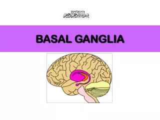
COMPONENTS • No generally accepted defination • Considered to include 1.corpus striatum﴾ neostriatum﴿ -putamen and caudate nucleus 2.claustrum 3.substantia nigra: pars compacta and pars reticularis 4.globus pallides 5. subthalamic nucleus
Cont’d • Play a major role in the control of posture and movement • Straitum- receptive component • Globus pallidus – its internal seg’t, output str. • Subthalamic – input from cer. cortex and reciprocal connection with G.P • Substantia nigra- pars compacta: contain dopamine -pars reticulata :continuation of internal segment of globus pallidus
connections • Striatum receives three main inputs -excitatory input from cerebral cortex -excitatory input from intralaminar thalamic nuclei -modulatory input from substantia nigra PC. • Striatal efferents -inhibitory GABAergic cells that project to the G.p and substantia nigra
Cont’d • Internal segment of G.P -project to three main targets -to thalamus -tosuperior colliculus -pedenculopontine nuclei of reticular form • Output of B.G affects both corticospinal and brainstem motor pathways
Lesions of B.G • Subthalamic nuc.- contralat. Hemiballismuss • Caudate nucleus- contralat. Choreoathetosis • Globus pallidus- -unilat.: contralat.hemidystonia ,hemiparkin. or tremor -bilat.:dystonia ,parkisonism abulia, akinesia • Substantia nigra -parkinsonism
Cont’d • Two main types of disorders of B.G ·hyperkinetic﴾ dyskinetic ﴿ disorders -when dopaminergic mechs.exagerated ·hypokinetic ﴾akinetic ﴿disorders -when dopaminergic mechs. impaired
Dyskinesias • Chorea • Tardive and orofacial dyskinesias • Ballismus • Athetosis, dystonia, torticollis • Myoclonus • Tics • tremor
Hypokinetic disorders • Parkinsonism • Corticobasal degeneration • Progressive supranuclear palsy • Multiple systems atrophy