Download
1 / 30
300 likes | 303 Views
This chapter provides an introduction to animal structure and function, focusing on the different types of tissues and the nervous system. It covers epithelial, connective, and nervous tissues, as well as the autonomic and central nervous systems. The chapter also explores the organization of the brain and the functions of its different hemispheres and lobes.

E N D
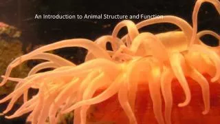
Chapter 40 An Introduction to Animal Structure and Function Chapter 48 Nervous System
Tissues: groups of cells with a common structure and function (4 types) Anatomy: structure Physiology: function • 1- Epithelial: outside of body and lines organs and cavities; held together by tight junctions • basement membrane: dense mat of extracellular matrix • mucous membrane (glandular epithelia) secrete mucus – digestive & respiratory • Simple: single layer of cells • Stratified: multiple tiers of cells • Cuboidal (like dice) • Columnar (like bricks on end) • Squamous (like floor tiles)
Tissues, II • 2- Connective: bind and support other tissues; extensive extracellular matrix; 3 kinds: • A-Collagenous fibers (collagen protein) B-Elastic fibers (elastin protein) C-Reticular fibers (thin branched collagen fibers) • Loose connective tissue: binds epithelia to underlying tissue; holds organs • 1-Fibroblasts- secretes extracellular proteins 2-Macrophages- amoeboid WBC’s; phagocytosis • 3-Adipose tissue- fat storage; insulation • Fibrous connective tissue: Dense-large number of collagenous cells. Parallel bundles of cells • 1-Tendons- muscles to bones 2-Ligaments- bones to bones; joints (BOBOLI) • Cartilage: collagen in a rubbery matrix (chondroitin); flexible support • Bone: mineralized tissue by osteoblasts • Blood: liquid plasma matrix; erythrocytes (RBC’s) carry O2; leukocytes (WBC’s) immunity
Tissues, III • 3-Nervous: senses stimuli and transmits signals from 1 part of the animal to another • Neuron: functional unit that transmits impulses • Dendrites: transmit impulses from tips to rest of neuron • Axons: transmit impulses toward another neuron or effector
Autonomic System • Two divisions: • sympathetic • Parasympatheitic • Control involuntary functions • heartbeat • blood pressure • respiration • perspiration • digestion • Can be influenced by thought and emotion
CENTRAL NERVOUS SYSTEM SYMPATHETIC Brain Dilates pupil Stimulates salivation Salivary glands Relaxes bronchi Spinal cord Lungs Accelerates heartbeat Heart Inhibits activity Stomach Pancreas Stimulates glucose Liver Adrenal gland Secretion of adrenaline, nonadrenaline Kidney Relaxes bladder Sympathetic ganglia Stimulates ejaculation in male Sympathetic • “ Fight or flight” response • Release adrenaline and noradrenaline • Increases heart rate and blood pressure • Increases blood flow to skeletal muscles • Inhibits digestive functions
CENTRAL NERVOUS SYSTEM PARASYMPATHETIC Brain Contracts pupil Stimulates salivation Constricts bronchi Spinal cord Slows heartbeat Stimulates activity Stimulates gallbladder Gallbladder Contracts bladder Stimulates erection of sex organs Parasympathetic • “ Rest and digest ” system • Calms body to conserve and maintain energy • Lowers heartbeat, breathing rate, blood pressure
Brain Spinal Cord Central Nervous System • Brain and Spinal Cord
Corpus Callosum Right Hemisphere Left Hemisphere Brain has 2 Hemispheres • Left & Right sides are separate • Corpus Callosum : major pathway between hemispheres • Some functions are ‘lateralized’ • language on left • math, music on right • Lateralization is never 100%
Frontal Parietal Occipital Temporal Each hemisphere is divided into 4 lobes
Left visual field Right visual field Optic nerves Left Visual Cortex Corpus Callosum Right Visual Cortex Sensory Information sent to opposite hemisphere • Principle is Contralateral Organization • Sensory data crosses over in pathways leading to the cortex • Visual Crossover • left visual field to right hemisphere • right field to left • Other senses similar
Motor Cortex Somatosensory Cortex Contralateral Motor Control • Movements controled by motor area • Right hemisphere controls left side of body • Left hemisphere controls right side • Motor nerves cross sides in spinal cord
Medial surface of right hemisphere Corpus Callosum Corpus Callosum • Major ( but not only) pathway between sides • Connects comparable structures on each side • Permits data received on one side to be processed in both hemispheres • Aids motor coordination of left and right side
Occipital Lobe • Input from Optic nerve • Contains primary visual cortex • most is on surface inside central fissure • Outputs to parietal and temporal lobes Occipital Lobe Visual Lobe
Auditory Cortex Temporal Lobe Temporal Lobe Temporal Lobe • Contains primary auditory cortex • Inputs are auditory, visual patterns • speech recognition • face recognition • word recognition • memory formation • Outputs to limbic System, basal Ganglia, and brainstem
Somatosensory Cortex Parietal Lobe Parietal Lobe • Inputs from multiple senses • contains primary somatosensory cortex • borders visual & auditory cortex • Outputs to Frontal lobe • hand-eye coordination • eye movements • attention
Frontal Lobe Working Memory Motor Cortex Motor Cortex Motor Cortex Broca’s Area Frontal Lobe • Contains primary motor cortex • No direct sensory input • Important planning and sequencing areas • Broca’s area for speech • Prefrontal area for working memory
Tissues, IV • 4- Muscle: capable of contracting when stimulated by nerve impulses; myofibrils composed of proteins actin and myosin; 3 types: • A- Skeletal: voluntary movement (striated) • B- Cardiac: contractile wall of heart (branched striated) • C- Smooth: involuntary activities (no striations)
Organ: organization of tissues Mesentaries: suspension of organs (connective tissue) Thoracic cavity (lungs and heart) Abdominal cavity (intestines) Diaphragm (respiration) Organ systems…... Digestive-food processing Circulatory-internal distribution Respiratory-gas exchange Immune/Lymphatic-defense Excretory-waste disposal; osmoregulation Endocrine-coordination of body activities (homeostasis) Reproductive-reproduction Nervous-detection of stimuli Integumentary-protection Skeletal-support; protection Muscular-movement; locomotion Organ systems
Internal regulation • Interstitial fluid: internal fluid environment of vertebrates; exchanges nutrients and wastes • Homeostasis: “steady state” or internal balance • Negative feedback: change in a physiological variable that is being monitored triggers a response that counteracts the initial fluctuation; i.e., body temperature • Positive feedback: physiological control mechanism in which a change in some variable triggers mechanisms that amplify the change; i.e., uterine contractions at childbirth
Metabolism: sum of all energy-requiring biochemical reactions • Catabolic processes of cellular respiration • Calorie; kilocalorie/C • Endotherms: bodies warmed by metabolic heat • Ectotherms: bodies warmed by environment • Basal Metabolic Rate (BMR): minimal rate for powering basic functions of life (endotherms) • Standard Metabolic Rate (SMR): minimal rate powering basic functions of life at a given temperature (ectotherms)