Download
1 / 61
640 likes | 858 Views
Ankle and Lower Leg Rehabilitation. Figure 15-1. Functional Anatomy. Talocrural Joint Articulation of distal end of the Tibia and Fibula with superior, medial and lateral aspect of Talus Referred to as ankle mortise 2 movements Ankle Dorsi-flexion and ankle Plantar-Flexion

E N D
Functional Anatomy • Talocrural Joint • Articulation of distal end of the Tibia and Fibula with superior, medial and lateral aspect of Talus • Referred to as ankle mortise • 2 movements • Ankle Dorsi-flexion and ankle Plantar-Flexion • 20 degrees DF and 50 degrees PF • Normal gait requires 20 deg. PF and 10deg. DF
Functional Anatomy • Talocrural joint ligaments • Lateral: anterior talofibular ligament (ATFL), Calcaneofibular Ligament (CFL), Posterior talofibular ligament (PTFL) • Medial: Deltoid Ligament; anterior, middle and posterior bands • Anterior & Posterior Tibiofibular ligament • Distal portion of interosseous membrane
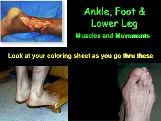
Talocrural muscles • Posterior to lateral malleolous create plantar flexion and toe flexion • Superficial: gastrocnemius • Middle: soleus & plantaris • Deep: posterior tibialis, flexor digitorum longus, flexor hallucis longus • Anterior muscles will dorsiflex the ankle and extend the toes • Ext. halicus longus, tibialis anterior, extensor digitorum, peroneal tertius
Subtalar joint • Articulation of calcaneus and talus • Pronation and supination • Occur in 3 planes simultaneously • Supination: Foot moves into plantar flexion, adduction, and inversion • Pronation: Foot moves into abduction, dorsiflexion and eversion
Midtarsal joint • Calcaneocuboid joint (CC) • Talonavicular joint (TN) • Depend on ligamentous and muscle tension to maintain position and integrity • Directly related to position of subtalar joint • If pronated, TN & CC become hypermobile • If supinated TN & CC become hypomobile
MT joint during pronation • Hypermobile 1st ray and increase pressure on other metatarsals • Peroneal tendon unable to stabilize 1st ray because mechanical advantage lost at cuboid pulley • Also hypermobility at articulation between 1st metatarsal and 1st cuneiform
Functional Anatomy • MT joint during supination • Less surface area between tarsal articulation=less movement=hypomobility • Foot rigid and tight • More weight and stress placed on 1st and 5th metatarsal because of less mobility at 1st ray
Functional Anatomy • Ankle more unstable in plantar flexion • Shape of talus: Wider anteriorly and more narrow posteriorly • In Dorsi flexion talus gripped tightly in talocrural joint • In plantar flexion less stable because narrow aspect of talus exposed • Also less stable with inversion • Distal end of tibia doesn’t extend as far as distal end of fibula
Biomechanics of Normal Gait • 2 phases: stance or support phase & swing or recovery phase • Stance: initial contact at heel strike and ends at toe off • Swing: time immediately after toe off, leg moved from behind body to a position in front of body in preparation of heel strike
Foot at stance phase • Shock absorber to impact forces at heel strike and adapt to uneven surface • At push off functions as rigid lever to transmit explosive force • Lateral aspect of calcaneus with subtalar joint in supination to forefoot contact on medial surface of foot and subtalar joint pronation • Pronation distributes forces to many structures
Foot begins to re-supinate and returns subtalar joint to neutral ay 70 to 90 % of support phase • Foot becomes rigid and stable to allow greater amount of force at push off
Assessing the Lower Leg and Ankle • History • Past history • Mechanism of injury • When does it hurt? • Type of, quality of, duration of pain? • Sounds or feelings? • How long were you disabled? • Swelling? • Previous treatments?
Observations • Postural deviations? • Genu valgum or varum? • Is there difficulty with walking? • Deformities, asymmetries or swelling? • Color and texture of skin, heat, redness? • Patient in obvious pain? • Is range of motion normal? • Palpation • Begin with bony landmarks and progress to soft tissue • Attempt to locate areas of deformity, swelling and localized tenderness
Ankle Stability Tests • Anterior drawer test • Used to determine damage to anterior talofibular ligament primarily and other lateral ligament secondarily • A positive test occurs when foot slides forward and/or makes a clunking sound as it reaches the end point • Talar tilt test • Performed to determine extent of inversion or eversion injuries • With foot at 90 degrees calcaneus is inverted and excessive motion indicates injury to calcaneofibular ligament and possibly the anterior and posterior talofibular ligaments • If the calcaneus is everted, the deltoid ligament is tested
Bump Test Talar Tilt Test Anterior Drawer Test
Functional Tests • While weight bearing the following should be performed • Walk on toes (plantar flexion) • Walk on heels (dorsiflexion) • Hops on injured ankle • Start and stop running • Change direction rapidly • Run figure eights
Footwear • Can be an important factor in reducing injury • Shoes should not be used in activities they were not made for • Preventive Taping and Orthoses • Tape can provide some prophylactic protection • However, improperly applied tape can disrupt normal biomechanical function and cause injury • Lace-up braces have even been found to be effective in controlling ankle motion
Neuromuscular Control Training • Can be enhanced by training in controlled activities on uneven surfaces or a balance board Figure 15-5 & 6
PHASE I • Decrease pain and swelling • PRICE • Modalities: pulsed ultrasound, electrical stimulation (Interferential, High Volt) • Massage • Pain-free AROM exercises
Phase II-ROM • Increase ROM: • AROM, PROM exercises • Progress to weight bearing ROM ex. • Maintain CV fitness • Maintain Core Stability • Restore Balance and proprioception • Double leg and single leg balance progression • Continue to assist healing process and pain management
Phase III-Strengthening • Continue ROM exercises • Continue to assist healing process and pain management • Continue and progress CV fitness • Continue and progress Core stability • Evaluate and treat other biomechanical deficiencies • Begin strengthening programs for foot and ankle as well as entire lower kinetic chain • Progress to functional activities and plyometrics
Phase IV • Continue all of Phase III • Add sport specific movement exercises • Rehab should be equally, if not more difficult than their practice for their sport • Running progression • Speed and agility • Sport specific movement • Goal of Phase IV is return to their sport
Phase V-Maintenance • Continue to monitor and rehabilitate athlete through their return to activity • Observe for setbacks or decrease in performance • Ensure activity and movement is coordinated and unconscious • Athlete should not be limited at all by their injury
Recognition and Management of Injuries to the Ankle • Ankle Injuries: Sprains • Single most common injury in athletics caused by sudden inversion or eversion moments • Inversion Sprains • Most common and result in injury to the lateral ligaments • Anterior talofibular ligament is injured with inversion, plantar flexion and internal rotation • Occasionally the force is great enough for an avulsion fracture to occur w/ the lateral malleolus
Severity of sprains is graded (1-3) • With inversion sprains the foot is forcefully inverted or occurs when the foot comes into contact w/ uneven surfaces
Eversion Ankle Sprains -(Represent 5-10% of all ankle sprains) • Etiology • Bony protection and ligament strength decreases likelihood of injury • Eversion force resulting in damage to deltoid and possibly fx of the fibula • Deltoid can also be impinged and contused with inversion sprains
Syndesmotic Sprain • Etiology • Injury to the distal tibiofemoral joint (anterior/posterior tibiofibular ligament) • Torn w/ increased external rotation or dorsiflexion • Injured in conjunction w/ medial and lateral ligaments • May require extensive period of time in order to return to play Figure 15-13
Graded Ankle Sprains • Signs of Injury • Grade 1 • Mild pain and disability; weight bearing is minimally impaired; point tenderness over ligaments and no laxity • Grade 2 • Feel or hear pop or snap; moderate pain w/ difficulty bearing weight; tenderness and edema • Positive talar tilt and anterior drawer tests • Possible tearing of the anterior talofibular and calcaneofibular ligaments • Grade 3 • Severe pain, swelling, hemarthrosis, discoloration • Unable to bear weight • Positive talar tilt and anterior drawer • Instability due to complete ligamentous rupture
Care • Must manage pain and swelling • Apply horseshoe-shaped foam pad for focal compression • Apply wet compression wrap to facilitate passage of cold from ice packs surrounding ankle • Apply ice for 20 minutes and repeat every hour for 24 hours • Continue to apply ice over the course of the next 3 days • Keep foot elevated as much as possible • Avoid weight bearing for at least 24 hours • Begin weight bearing as soon as tolerated • Return to participation should be gradual and dictated by healing process
Ankle Fractures/Dislocations • Cause of Injury • Number of mechanisms – often similar to those seen in ankle sprains • Signs of Injury • Swelling and pain may be extreme with possible deformity • Care • Splint and refer to physician for X-ray and examination • RICE to control hemorrhaging and swelling • Once swelling is reduced, a walking cast or brace may be applied, w/ immobilization lasting 6-8 weeks • Rehabilitation is similar to that of ankle sprains once range of motion is normal
Tendinitis • Cause of Injury • Singular cause or collection of mechanisms • Footwear, mechanics, trauma, overuse, limited flexibility • Signs of Injury • Pain & inflammation • Crepitus • Pain with AROM & PROM • Care • Rest, NSAIDs, modalities • Orthotics for foot mechanic
Tibial and Fibular Fractures • Cause of Injury • Result of direct blow or indirect trauma • Fibular fractures seen with tibial fractures or as the result of direct trauma • Signs of Injury • Pain, swelling, soft tissue insult • Leg will appear hard and swollen (Volkman’s contracture) • Deformity – may be open or closed • Care • Immediate treatment should include splinting to immobilize and ice, followed by medical referral • Restricted weight bearing for weeks/months depending on severity
Stress Fracture of Tibia or Fibula • Cause of Injury • Common overuse condition, particularly in those with structural and biomechanical insufficiencies • Result of repetitive loading during training and conditioning • Signs of Injury • Pain with activity • Pain more intense after exercise than before • Point tenderness; difficult to discern bone and soft tissue pain • Bone scan results (stress fracture vs. periostitis)
Care • Eliminate offending activity • Discontinue stress inducing activity 14 days • Use crutch for walking • Weight bearing may return when pain subsides • After pain free for 2 weeks athlete can gradually return to activity • Biomechanics must be addressed