Download
1 / 14
150 likes | 244 Views
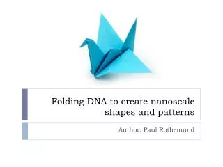
Paul Rothemund , Departments of Computer Science and Computation & Neural Systems, California Institute of Technology. Folding DNA to create nanoscale shapes and patterns. Jerzy Szablowski 20.309 : Biological Instrumentation and Measurement Fall 2008. Available methods:

E N D
Paul Rothemund, Departments of Computer Science and Computation & Neural Systems, California Institute of Technology Folding DNA to create nanoscale shapes and patterns • JerzySzablowski • 20.309: Biological Instrumentation and Measurement • Fall 2008
Available methods: • Atomic force microscopy • Scanning tunneling microscopy • micro-contact printing • Lithography • Self-assembly Nanopatterning Fabrication of sub-micron structures allowing for arbitrary positioning of molecules and, in some cases, single atoms. (“Top-bottom” method) Source: IBM, 1990 (“Bottom-up” method) Rothemund, 2006 Zhang, 2003
Self assembly using DNA • Can assembly multiple structures at a time. • Can occur in less stringent conditions than “top-down” methods (AFM, STM…). • Does not require expensive equipment • Complexity of patterns formed is not as high as in the other cases. Some exciting studies emerged: Chen and Seeman, 1991 Rothemund, 2006 Nanowire mesh, by Yan et al,2003 These approaches very sensitive to DNA concentrations, and require multiple reactions and purification steps. Yet patterns complexity doesn’t match other methods of nanofabrication. Is it worth it?
How to build an arbitrary 2-D pattern with 6-nm resolution out of DNA. Draw out a shape you would like to obtain, determine the desired size. Sketch a single molecule of DNA through the shape, as in the picture below. Proceed with a custom written software. (Rothemund, 2006)
Computer aided design of DNA assemblies Design primers binding to two distant sites of the DNA molecules: this will fold DNA into a desired structure. It might be necessary to introduce additional bridges that will stabilize the structure. 6nm pixel Synthesize linkers and scaffold, add togather in a tube and cool down from 95C to 20C at 1C per minute.
Results All shapes based on a single M13mp18 DNA molecule. Containing up to 273 linkers. Many of the structures are assembled properly. Regardless of stoichiometry. • Based on probability • Stronger structures – with lower energy are more likely to assemble. • Mechanic disruption: inserting the AFM tip can destroy the DNA structures. • Sometimes also: AFM imaging artifact. 100 nm 100 nm 100 nm
Assembling higher order structures Structures can be assembled by adding “staples” on the side of an assembled molecule. Staples are put in on the rim which previously held its structure: Many other structures can be made! Both contained and of unlimited size. 100nm Scheme. Real result
Implanting the “pixels” • Brighter specks on the image represent labeled DNA. Up to 200 pixels can be placed. • Labels are pieces of DNA and anything that is attached to it, such as: - biotin, fluorophores… proteins?
Applications • Unlimited: each pixel can be a specific aptamerbinding a protein or a small molecule. Biotin also can serve as a very efficient adapter for binding. We could place molecules arbitrarily in space. Some ideas include: • Nanobreadbord: with nanowires, gold nanoparticles, maybe enzymes, fluorophores… • Small “catalyzing centers”, where molecules are patterned in a way favoring certain chemical reactions. • nano-Microarrays? Future: 3D structures, greater number of pixels.
Summary • “DNA origami”, allows for folding a DNA molecule into a highly predictable two-dimensional shape with 6nm resolution. • It requires “stitching” various parts of the molecule by short complementary primers (staples). • All these molecules can be implanted with DNA adapters (pixels) that can bind range of molecules • But sometimes only a fraction of molecules fold properly.
References and questions References Rothermund, P. W. K. Folding DNA to create nanoscale shapes and patterns. Nature 440, 298–302 (2006). Yan, H., Park, S. H., Finkelstein, G., Reif, J. H. & LaBean, T. H. DNA-templated self-assembly of protein arrays and highly conductive nanowires. Science 301,1882–-1884 (2003). Zhang, S. Fabrication of novel biomaterials through molecular self-assembly. Nat. Biotechnol.21, 1171–1178 (2003). Chen J, Seemen, N.C. The synthesis from DNA of a molecule with the connectivity of a cube. Nature, 350, 631-633 (1991) Questions ?
What about twisting DNA? Other steric hindrances? • It does twist. To avoid deleterious strain, staples are placed either facing upwards, or downwards, which in the longer run reduces the strain. • It also bends because of the DNA stiffness and curving on the edges. That’s why AFM images show
Would not M13 genome dimerize, leaving no space for staples? • Yes it would, but apparently thanks to very high concentration of staples and conditions of folding – it rarely does. Secondary structure of M13 genome, from supplementary material.