Download
1 / 47
470 likes | 473 Views
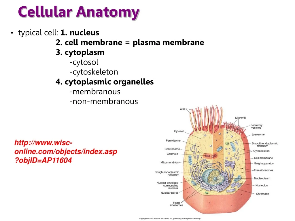
Cellular Anatomy. typical cell: 1. nucleus 2. cell membrane = plasma membrane 3. cytoplasm -cytosol -cytoskeleton 4. cytoplasmic organelles -membranous -non-membranous. http://www.wisc-online.com/objects/index.asp?objID=AP11604. The Typical Cell.

E N D
Cellular Anatomy • typical cell: 1. nucleus 2. cell membrane = plasma membrane 3. cytoplasm -cytosol -cytoskeleton 4. cytoplasmic organelles -membranous -non-membranous http://www.wisc-online.com/objects/index.asp?objID=AP11604
The Typical Cell TEM of a typical eukaryotic cell
The Nucleus: Information Central Nucleus Nucleolus Chromatin • nucleus contains most of the cell’s genes and is usually the most conspicuous organelle • bound by a double, phospholipid membrane = nuclear membrane • each membrane is a phospholipid bilayer • the nuclear envelope connects to the endoplasmic reticulum (protein synthesis) Nuclear envelope: Inner membrane Outer membrane 0.25 m Nuclear pore • nuclear membrane - contains nuclear pores = channels of over 100 proteins • allows entry and exit of materials e.g. mRNA Rough ER Porecomplex Pore complexes (TEM) Ribosome Close-upof nuclearenvelope Chromatin
Nucleus is comprised of the following • 1. Nucleoplasm • 2. Nucleolus • 3. Chromatin
A. Nucleoplasm: • specialized fluid of the nucleus • suspends the DNA in its form as chromatin • contains a network of filaments = nuclear matrix or nuclear lamina • matrix gives the nucleus its shape • matrix helps organize the chromatin • also helps in DNA replication & transcription 1 m Nuclear lamina (TEM)
B. Nucleolus: • “little nucleus” • dense body DNA, RNA and protein with no defining membrane • site of ribosome production and rRNA synthesis (via transcription) • site for the assembly of rRNA with the two protein subunits of the ribosome • two ribosome subunits migrate out through nuclear pores – for assembly in the cytoplasm • nucleolus can be very prominent in cells that produce high amounts of proteins (e.g. neurons)
C. Chromatin: • loosely coiled fibers of DNA wrapped around proteins = histones • found only in eukaryotic cells • Function of Chromatin: • to package DNA into a smaller volume to fit in the cell • to strengthen the DNA to allow mitosis • to prevent DNA damage • to control gene expression and DNA replication
C. Chromatin: • described as a “beads on a string” model histone - the histone DNA complex is called a nucleosome • DNA in between nucleosomes is called “linker DNA” – 50bp • the entire complex is known as the 10nm fiber or euchromatin
Chromatin Organization • -10nm fiber = euchromatin • 10nm fiber is packed further into a 30nm fiber = heterochromatin • compacted further by forming “loops” • eventually condenses into a chromosome DNA Nucleosome Euchromatin Heterochromatin Further looping and condensing Chromosome
organization of chromatin depends on the cell cycle – i.e. the stage of cell replication • during interphase of the cell cycle - two forms of chromatin: euchromatin and heterochromatin (more condensed form of chromatin) • with cell replication - heterochromatin condenses even more -> chromosomes • condensing of chromatin through its various forms – regulated by specific proteins • e.g. protein called condensin • chromatin arrangement is critical to DNA function • more condensed the less access the molecular “machinery” of gene expression has • human karyotype
Histones • complex of small, basic proteins • core of 4 proteins: H2A, H2B, H3, H4 • 2 complexes = 8 total proteins per histone • DNA wraps around this core = 10nm fiber (euchromatin) • plus 2 linker histones H1, H5 • linkers interact with the DNA and pack it into a thicker 30nm fiber (heterochromatin) • many histone amino acids are positively charged • so DNA-histone interaction is just an attraction between negatively charged DNA and positively charged histones • not dependent upon DNA sequence • proteins of the histone make ionic bonds to the acidic sugar-PO4 backbone of the DNA helix • bonds between histone and DNA can be modified by enzymes • e.g. methylation, phosphorylation • this modification can change the interaction between the DNA and the histone • can make the DNA either more or less accessible to the replication machinery • modifications can either make replication easier or harder
The Search for the Genetic Material • early in the 20th century, the identification of the molecules of inheritance loomed as a major challenge to biologists • when T. H. Morgan’s group showed that genes are located on chromosomes, the two components of chromosomes—DNA and protein—became candidates for the genetic material • key factor in determining the genetic material was choosing appropriate experimental organisms • role of DNA in heredity was first discovered by studying bacteria and the viruses that infect them
EXPERIMENT DNA as the source of genetic material Mixture ofheat-killedS cells andliving R cells Heat-killedS cells(control) Living S cells(control) Living R cells(control) • 1928 • Frederick Griffith – working on a vaccine for pneumonia (Streptococcus pneumonia) • worked with 2 strains – one harmless, one pathnogenic • killed the pathenogenic bacteria and mixed them with cultures of living, harmless bacteria • some of the harmless bacteria became pathenogenic • their progeny remained pathenogenic • DNA had to have been transferred from path. to harmless when he mixed the bacteria • called this transfer = transformation (assimilation of external by a cell) RESULTS Mouse dies Mouse healthy Mouse healthy Mouse dies Living S cells
DNA as the source of genetic material • transformation now defined as – change in genotype and phenotype due to the assimilation of external DNA by a cell • 1944 -Oswald Avery, Maclyn McCarty, and Colin MacLeod announced that the transforming substance was DNA • 14 year project!! • possible transforming agents: DNA, RNA or protein • broke open heat-killed bacteria and extracted their contents • treated each sample with chemicals that inactivated either DNA, RNA or protein • tested the modified samples for their ability to transform live, non-pathenogenic bacteria • only DNA worked in transforming harmless bacteria into pathogenic bacteria • lost this function once they were inactivated with chemicals • many biologists remained skeptical, mainly because little was known about DNA
Viral DNA Can Program Cells • more evidence for DNA as the genetic material - studies of viruses that infect bacteria • called bacteriophages(or phages) • 1952 - Alfred Hershey and Martha Chase performed experiments showing that DNA is the genetic material of a phage known as T2 • phage that normally infects E. coli strains • composed of DNA and protein • they designed an experiment showing that only the DNA enters an E. coli cell during infection
Viral DNA Can Program Cells DNA labelled Proteins labelled • labelled phages with radioactive sulfur to label proteins (batch #1) or phosphorus to label DNA (batch #2) • mixed the labelled phages with bacteria to cause infection • separated the bacteria from the phages and looked for what radioactive signal was in the isolated bacteria • confirmed and measured the presence of radioactive phosphorus in the transformed bacteria
Chargaff’s Rules • DNA is a polymer of nucleotides, each consisting of a nitrogenous base, a sugar, and a phosphate group • 1950 - Erwin Chargaff reported that DNA composition varies from one species to the next • BUT he also noticed two other things • these 2findings became known as Chargaff’s rules • 1. WHILE the base composition of DNA varies between species • 2. the number of A and T bases are equal and the number of G and C bases are equal in any species
Watson & Crick • 1950s – three groups working on the structure of DNA • 1. Linus Pauling – Cal Tech • 2. Maurice Wilkins and Rosalind Franklin – King’s College, London, UK • 3. American James Watson and Englishman Francis Crick – Cavendish Laboratory, Cambridge, UK • 1953- Watson and Crick constructed a model for DNA – two complementary strands that run anti-parallel to one another in a double helix
(b) Franklin’s X-ray diffractionphotograph of DNA Watson & Crick • based their work on Rosalind Franklin’s X-ray crystallography findings • X-rays bent as they passed through the strands of purified DNA • photo 51 – suggested a double helix, with the bases facing inward, making a full turn every 3.4nm, 10 base pairs every full turn • 1962 – Nobel prize – Watson, Crick and Wilkins (Franklin dies in 1958) (a) Rosalind Franklin
Sugar-phosphate backbone Nucleic acids 5 end 5C • two types: DNA, RNA • C,H,O,N,P • building blocks = nucleotides 3C Nucleoside • nucleotide: • 5 carbon sugar (pentose) • phosphate group (negative charge) located at the 5’ carbon • organic base located at the 1’carbon • sugar and the base is known as a nucleoside • bases: 5 types: adenine (A) • cytosine (C) • guanine (G) • thymine (T) • uracil (U) Nitrogenousbase 5C 1C Phosphategroup 3C Sugar(pentose) 5C 3C (b) Nucleotide 3 end (a) Polynucleotide, or nucleic acid
Naming nucleotides • nucleotide is conventionally named “based” on its base • adenine, guanine etc… • but the true name is a combination of the nucleoside and the number of phosphate groups • nucleoside= combination of a base and a sugar • e.g. adenine + sugar = adenosine • so “adenine” is really called • e.g. RNA - adenosine monophosphate (AMP) • e.g. DNA - deoxy-adenosine monophosphate (dAMP)
Pyrimidines Purines • There are two families of nitrogenous bases • 1. Pyrimidines(cytosine, thymine, and uracil)have a single six-membered ring • 2. Purines (adenine and guanine) have a six-membered ring fused to a five-membered ring Nitrogenous bases Cytosine (C) Uracil (U, in RNA) Thymine (T, in DNA) Sugars Deoxyribose (in DNA) Ribose (in RNA) Adenine (A) Guanine (G) (c) Nucleoside components
DNA is double stranded: The DNA double helix • DNA strand is known as a polynucleotide chain • 2 sugars of adjacent nucleotides are joined by a phosphodiester bond • the phosphodiester bond can only form between a 3’OH “down” to a 5’PO4 group • so that the DNA chain grows in a 5’ to 3’ direction
-the DNA strand must “grow” in a specific direction because of the nature of the phosphodiester bond -this direction is called 5’ to 3’ because one end is a 5’phosphate group, the other end is a 3’ OH group -the complementary strand also grows this way = two anti-parallel strands of DNA (opposite directions of growth)
Why 5’ to 3’?? • the DNA chain grows in a 5’ to 3’ direction • why can’t you form a phosphodiester bond from 3’ to 5’?? • the first thing that happens is the bases pair up with each other • THEN the phosphodiester bond forms • the incoming nucleotide is in itstriphosphate form
Why 5’ to 3’?? • a magnesium ion is associated with the outer two phosphate groups • this weakens the phosphate bonds between these groups • and allows for a successful nucleophilic attack by the 3’OH group in the nucleotide that is already bound to the DNA chain • results in the loss of the outer 2 phosphate groups (due to a shift in electrons) and the formation of a 3’-5’ phosphodiester bond Nucleophilic attack Mg2+
single DNA chain is 2.2 to 2.6nm (22 to 26 Angstroms) wide – each nucleotide is 3.3A long • DNA exists as a double helix – backbone made of alternating sugars and phosphates from the nucleotide • bases are inside the helix – away from the aqueous environment of the cell • helix makes 1 full turn every 3.4nm • ~10bp every turn • so one base is 0.34nm wide • helix is a right hand twist • clockwise twist
2 DNA strands are held together by Hydrogen bonds between the bases • not very strong, so DNA helix is easily separated by high heat, salt, mechanical forces etc… • this is known as denaturation • Watson and Crick found that the helix was uniform in its diameter • proposed that the base pairing had to be consistent throughout the helix • proposed = DNA held together by complementary base pairs • the base pairing rule is purine-pyrimidine • purine-purine pairing would produce a bulge in the helix • pyrimidine-pyrimidine pairing would produce a “dent” • A to T – two H bonds • C to G - three H bonds • C-G pairings are stronger – DNA is harder to separate in those regions (“GC rich”)
helix also exhibits additional levels of coiling = supercoiling • DNA can either supercoil in the direction of the helix (positive) or in the opposite direction (negative) • degree of supercoiling can determine the strength of the interaction between the two DNA chains
Naming the DNA strands • DNA is a double helix of complementary sequence • to simplify things we call one strand “sense”the other “anti-sense” • sense strand – has same sequence as the mRNA strand that will be translated into a protein • anti-sense strand – template used to create this mRNA • when written out – the sense strand is the top strand – with the 5’PO4 on the left and the 3’OH on the right
-double stranded DNA helix is unwound into individual strands -enzyme = DNA polymerase attaches to each strand -polymerase moves along the DNA strand and attaches the complementary base e.g. C is read – attaches a G
Models of DNA replication • conservative: means that the replicated DNA helices are entirely parental or daughter • semi-conservative: means that the replicated DNA helices are 50% parental DNA and 50% daughter DNA • dispersive: means that the replicated DNA helices are a mixture of parental and daughter DNA
Replication is Semi-conservative • labelled bacterial DNA first with a heavy 15N isotope to label the parental strands • replication - added a labelled a lighter 14N isotope that would label the replicated DNA daughter strands • continued to label bacteria with the lighter isotope for another round of replication • isolated the DNA & based on their “weight” they could tell which DNA strands had the 15N label or the 14N label • found that after the 1st replication – hybrids of both 15/14N strands • after the second replication – mixture of hybrid DNA and light 14N DNA helices • only way to explain this = Semi-conservative replication
DNA replication – the players • DNA polymerase III= reads the DNA strand and lays down a complementary base to create a complementary “daughter” strand of new DNA • Helicase/dnaB= enzyme that “melts” or unzips the double-helix of the parental DNA • single stranded binding proteins/SSBs– hold the unwound DNA helix “open” – allows for the action of the replication machinery • RNA polymerase (Primase/dnaG)= creates a small RNA “oligo” primer which binds onto the DNA template and allows the DNA polymerase III to attach to the DNA template • DNA ligase= joins up the fragments of DNA made during replication
DNA polymerase • polymerase = enzyme that catalyzes the addition of bonds between two nucleotides of either DNA or RNA • catalyzes the bond between bases • also catalyzes the phosphodiester bond within the backbone • in bacteria – three polymerases that act in replication • replication polymerase = DNA polymerase III • DNA polymerase I – removes RNA nucleotides found in the primer(exonuclease activity) and fills in the gaps • also corrects mistakes made upon replication = “proof-reading” • DNA polymerase II – proof reading • in eukaryotic cells – 11 polymerases !! • same idea as bacterial replication
Problems with Replication • first problem: DNA is a helix • DNA polymerases are unable to melt duplex DNA in order to separate the two strands that are to be copied • solution: binding of a “helicase”enzyme unwinds the two strands • second problem: unwinding the DNA produces supercoiling in the regions ahead • solution: action of topoisomerasesto “nick” the DNA helix, unwind the supercoils and connect the strand back together
Problems with Replication • another problem: the helix wants to re-form • solution: single-stranded binding proteins (SSBs)also required to prevent the DNA template from rewinding back up • still another problem: DNA polymerases cannot bind single stranded nucleic acids • unwinding the DNA gets rid of base pairing = Problem ! • DNA polymerase III requires a “primer” to elongate off of • solution: enzyme called a “primase”makes a small piece of RNA that binds to the DNA strand and acts as a primer for the DNA polymerase – creates a temporary double stranded structure of RNA and DNA
Problems with the DNA polymerase • Biggest Problem: The two strands in the DNA duplex are opposite in chemical polarity • DNA polymerase can only form a phosphodiester bond between the 5’PO4 of an incoming new nucleotide and a 3’OH of a nucleotide already base paired to the DNA template • this means that new DNA strands can grow only in the 5 to 3 direction • this is okay when replicating the anti-sense strand because the complementary daughter strand that is being created will grow in the 5’ to 3’ direction • but there is a problem replicating the sense strand 5’ 3’ growing daughter strand DNA poly 3’ 5’ growing daughter strand
DNA replication • replication starts at specific sequences of DNA = origins of replication • called oriC in bacteria • ~240bp sequence – containing repetitive sequences – rich in As and Ts • also found in viruses • multiple origins are found in eukaryotic chromosomes • oriC is recognized by a protein complex that docks onto the oriC • this complex is comprised of at least five protein complexes –the helicase, the primase, DNA polymerase, clamp loading protein, infrastructure proteins that ‘tether’ the two DNA polymerases to each other
DNA replication • oriC is recognized by the helicase that unwinds the DNA • “melts” the DNA • forms a replication “bubble” • comprised of two replication forks • the forks are held open by SSBs • the helicase can actually act as a molecular “brake” that controls how fast replication happens the helicase/primase/DNA polymerase moves along the parental DNA strand in the 3’ to 5’ direction
DNA replication: The Leading strand • REMEMBER – a complex of proteins binds at the oriC and moves along the parental DNA in the 3’ to 5’ direction • because of this one strand of parental DNA is able to be replicated continuously without any problems • new daughter strand grows in the 5’ to 3’ direction • the DNA strand that is made continuously = Leading Strand • the leading strand is uses the anti-sense strand of parental DNA as its template • the leading strand also grows in the same direction as the replication fork Replication fork direction
DNA replication: The Lagging strand • but since the second strand of DNA is in the opposite direction – it cannot be replicated continuously • this would require the daughter strand be made in the 3’ to 5’ direction • solution – sense strand is replicated in “chunks” • this daughter DNA strand being made is called the Lagging strand • ‘chunks’ are called Okazaki fragments – 1000 to 2000 bps • (Reiji Okazaki) • as the helicase unwinds the parental duplex, primase creates multiple primers along the sense parent strand • DNA polymerase “clamps” onto the 3’ end of this DNA-RNA hybrid duplex and makes an Okazaki fragment
DNA polymerase III – 2 large complexes tethered together • core polymerase – three subunits: • alpha – active site for nucleotide addition – polymerase action • epsilon – exonuclease activity – part of the enzyme that removes incorrectly added nucleotides – “proof-reading” • can only back-up one nucleotide and correct it • so DNA polymerase III has to “catch” its mistakes as it makes them • sigma – proof-reading • beta – forms a beta-clamp – forms a donut-like clamp around the parental DNA • the gamma subunit: loads the b-clamp onto the DNA strand at the RNA primers and takes the clamp back off • now known as the Clamp-loading Protein
The big picture!! • leading and lagging strands are made at the same time • two core DNA polymerases III bind at each fork and replicate the DNA in the same direction! • how is this possible?? • the parental/lagging strand is “looped” within the complex so that its orientation is the same as the parental/leading strand • IMPORTANT – the DNA polymerase III complex doesn’t move – the DNA template is “fed into” the complex and then fed back out • DNA polymerase III is anchored to the nuclear matrix SSBs DNA polymerase https://www.youtube.com/watch?v=TNKWgcFPHqw (topoisomerase) Helicase & Primase direction of polymerase movement (3’ to 5’)
The big picture animation • for the “big picture”: http://www.youtube.com/watch?v=-mtLXpgjHL0 Leading Daughter- Antisense parent Sense parent Beta clamp Anti- Sense parent alpha helicase primase alpha SSBs RNA primer #2 RNA primer #1 Okazaki Fragment #1 Beta clamp
Leading Daughter- Antisense parent Polymerase direction Sense parent Anti- Sense parent alpha RNA primer #3 helicase alpha primase SSBs RNA primer #2 Okazaki Fragment #2 RNA primer #1 Okazaki Fragment #1
Check out these animations!! • http://www.ncc.gmu.edu/dna/repanim.htm • http://www.johnkyrk.com/DNAreplication.html • http://www.bioteach.ubc.ca/TeachingResources/MolecularBiology/DNAReplication.swf • http://bioweb.uwlax.edu/GenWeb/Molecular/Theory/Replication/replicat.mov