Download
1 / 51
510 likes | 691 Views
Time Since Death. CHS Forensics. Crime scene. The body must first be checked for signs of life. The body must not be moved. Photographs need to be taken to record the scene. Crime scene.

E N D
Time Since Death CHS Forensics
Crime scene The body must first be checked for signs of life. The body must not be moved. Photographs need to be taken to record the scene.
Crime scene • Investigators can then search the victim including the clothing (colors, size, labels, presence of buttons, and trace materials) • Coverings should be placed over the hands.
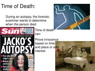
DEATH • Death is the extinction or cessation of life, but since life itself is difficult to define there is also a problem in defining death. • An added difficulty is that in nearly all circumstances human death is a process rather than an event.
TIME SINCE DEATH • All death certificates require an estimate of the time of death as well as a statement of the underlying cause of death. • If the death was witnessed then providing a time of death presents no difficulty, but in an un-witnessed death it can be problematic.
TIME SINCE DEATH Establishing the time of death is of assistance in any police investigation of a death, whether it was from natural or un-natural causes. Establishing the time of an assault and the time of death is critical in criminal proceedings in which there are legal issues of alibi and opportunity to commit the crime.
TIME SINCE DEATH • If an accused can prove that he was at some other place when the injury and death of the victim occurred then his innocence is implicit. • However, the time of injury, may be separated from the time of death by a significant survival period.
Sources of Evidence Look Away • Evidence for estimating the time of death may come from three sources: • Corporal evidence, present in the body. • Environmental and associated evidence, that is present in the vicinity of the body, • Anamnestic evidence, based on the deceased's ordinary habits, movements, and day to day activities.
Types of evidence used in finding T.O.D. • OCULAR CHANGES • ALGOR MORTIS • PALLOR MORTIS • RIGOR MORTIS • LIVOR MORTIS • POSTMORTEM DECOMPOSITION • GASTRIC CONTENTS • FORENSICS ENTOMOLOGY
Ocular changes Thin film appears over the cornea of opened eye within minutes of death (closed eyes- hours) • Corneal cloudiness (2-3 hours in open eyes and 24 hours in closed eyes) • Tache noire-blackish discoloration develops • No intraocular fluid after four days
ALGOR MORTIS BODY COOLING • Is the use of body temperature estimations to assess time of death • It is of some importance to note that this only applies to cool and temperate climates since in tropical regions there may be a minimal fall in body temperature post mortem
ALGOR MORTIS BODY COOLING • In some extreme climates, (e.g. central Australia) the body temperature may even rise after death.
ALGOR MORTIS BODY COOLING • The assessment is made on the basis of measurement of the body core temperature. • In practice either the temperature is measured intra-hepatic/sub-hepatic temperature.
Pallor Mortis • Defined as ‘Paleness of Death’. • Tone of the body. • Happens 15-20 minutes after death. • Happens due to lack of capillary circulation in the body. • Can not be used to determine time of death except if body is found still with color.
RIGOR MORTIS • Ordinarily, death is followed immediately by total muscular relaxation, succeeded in turn by generalized muscular stiffening - rigor mortis. • After a variable period of time rigor mortis passes off spontaneously to be followed by secondary muscular flaccidity.
RIGOR MORTIS Immediately following death-body is flaccid, followed by increasing rigidity due to lack of ATP and buildup of lactic acid Adenosine Triphosphate (ATP)-energy source produced in respiration in mitochondria of cells
RIGOR MORTIS • Generally rigor commences in 6 hours, takes another 6 to become fully established, remains for 12 hours and passes off during the succeeding 12 hours.
LIVOR MORTIS Lividety • Lividity is a dark purple discoloration of the skin resulting from the gravitational pooling of blood in the veins and capillary beds of the dependent parts of the body following cessation of the circulation.
LIVOR MORTIS Lividety • Lividity is present in all bodies, although it may be inconspicuous in some and thus escape notice. • Within about 30- 60 minutes of death the blood in most corpses, dead from natural or non-natural causes, becomes permanently incoagulable.
LIVOR MORTIS Lividety In deaths from carbon monoxide poisoning, it is classically described as "cherry red“ In cases where methaemoglobin is formed in the blood during life (e.g. potassium chlorate, nitrates, and aniline poisoning) it appears chocolate brown
LIVOR MORTIS Lividety In deaths from exposure to cold, it is bright pink, and a similar colouration is seen in bodies refrigerated very soon after death. Refrigeration of a body already displaying typical purple lividity will cause it to turn pink.
Gastric contents • If the last known meal is still present in the stomach of a corpse and the time of that meal is known, then it can give some general indication of the interval between the meal and death. • Digestion takes between ½ hour-6 hours depending on size and content of meal
Gastric contents • Affected by many factors: • Density-increase density-slower digestion • Drugs and alcohol-alcohol slows down digestion and narcotics speed up digestion • Medical Conditions-Diabetes delays digestion and shock causes content retention for days
Entomology • Insects will colonize a corpse if given the opportunity. • The most important flies whose larvae (maggots) feed on corpses belong to the groups Calliphoridaeor blow-flies and Sarcophagidaeor flesh-flies.
Calliphoridae Sarcophagidae
Entomology • Each part of the world has its own indigenous species of these flies. • While fly larvae feed on the corpse, beetles feed on the larvae, although some beetle groups will feed directly on the corpse. • Beetles appear on the corpse later than flies and are some of the last insects to colonise fragments of soft tissue remaining on skeletonised bodies
Stages of decomposition Fresh Stage Bloat Stage Active Decay Stage Advanced Decay Stage Dry Decay Stage
fresh stage The fresh stage of decomposition is generally described as the period between the moment of death and when the first signs of bloat are apparent. There are no outward signs of physical change, though internal bacteria have begun to digest organ tissues[.
fresh stage No odor is associated with the carcass. Early post-mortem changes, used by pathologists as medical markers for early post-mortem interval estimations; livor mortis, rigor mortis and algor mortis.
fresh stage The first insects to arrive at decomposing remains are usually Calliphoridae (blow flies), arrive within minutes of death or exposure, and deposit eggs within 1-3 hours. Flies of the families Sarcophagidae (flesh flies) and Muscidae are also common in this first stage of decomposition.
Bloat Stage The first visible sign of the bloat stage is a slight inflation of the abdomen and some blood bubbles at the nose. Activity of anaerobic bacteria in the abdomen create gases, which accumulate and results in abdominal bloating.
Bloat Stage A colour change is observed in the carcass flesh, along with the appearance of marbling. During the bloat stage the odor of putrefaction becomes noticeable.
Bloat Stage First species of Coleoptera arrive, Staphylinidae (rove beetles), Silphidae (carrion beetles) and Cleridae. These beetles are observed feeding on fly eggs and larvae. Beetle species from the families Histeridae may also be collected during this stage, and are often hidden beneath remains.
Active Decay Stage Adipocere Also known as "grave wax," adipocere (from the Latin, adipo for fat and cera for wax) is a grayish-white postmortem (after death) matter caused by fat decomposition, which results from hydrolysis and hydrogenation of the lipids (fatty cells) that compose subcutaneous (under the skin) fat tissues.
Active Decay Stage The beginning of active decay stage is marked by the deflation of the carcass as feeding Dipteran larvae pierce the skin and internal gases are released. During this stage the carcass has a characteristic wet appearance due to the liquefaction of tissues..
Active Decay Stage A strong odor of putrefaction is associated with the carcass. Feeding larvae of Calliphoridae flies are the dominant insect group at carcasses during the active decay stage.
Active Decay Stage At the beginning of the stage larvae are concentrated in natural orifices, which offer the least resistance to feeding. Towards later stages, when flesh has been removes from the head and orifices, larvae become more concentrated in the thoracic and abdominal cavities.
Advanced Decay Stage Most of the flesh is removed from the carcass during the advanced decay stage, though some flesh may remain in the abdominal cavity. Strong odors of decomposition begin to fade.
Advanced Decay Stage This stage marks the first mass migration of third instarcalliphorid larvae from the carcass Piophilidae larvae may also be collected at this stage. Few adult calliphoridae are attracted to carcasses in advanced decay.
Advanced Decay Stage Adult Dermestidae (skin beetles) arrive at the carcass; adult dermestid beetles may be common, whereas larval stages are not
Dry Decay The final stage of decomposition is dry remains. Very little remains of the carcass in this stage, mainly bones, cartilage and small bits of dried skin. There is little to no odor associated with remains.
Dry Decay The greatest number of species are reported to occur in the late decay and dry stages. The dry decay stage is characterized by the movement from previously dominant carrion fauna to new species.
Dry Decay The dermestid beetles, common in advanced decay, leave the carcass. Non-carrion insects that commonly arrive at remains in dry decay are centipedes, millipedes, isopods, snails and cockroaches.