Download
1 / 48

E N D
Chapter 7. Cartilage Chapter 7 Cartilage Prof Abdel-Majeed Safer Histology & Hisctochemistry
Cartilage: IntroductionCartilage is characterized by an extracellular matrix (ECM) enriched with glycosaminoglycans and proteoglycans, macromolecules that interact with collagen and elastic fibers. Variations in the composition of these matrix components produce three types of cartilage adapted to local biomechanical needs. Chapter 7 Cartilage Prof Abdel-Majeed Safer Histology & Hisctochemistry
Cartilage is a specialized form of connective tissue in which the firm consistency of the ECM allows the tissue to bear mechanical stresses without permanent distortion. In the respiratory system cartilage forms a framework supporting soft tissues. Because it is smooth-surfaced and resilient, cartilage provides a shock-absorbing and sliding area for joints and facilitates bone movements. Cartilage is essential for the development and growth of long bones, both before and after birth. Chapter 7 Cartilage Prof Abdel-Majeed Safer Histology & Hisctochemistry
Cartilage consists of cells (chondrocytes ), An extensive extracellular matrix composed of fibers and ground substance. Chondrocytessynthesize and secrete the ECM and the cells themselves are located in matrix cavities called lacunae.Collagen, hyaluronic acid, proteoglycans, and small amounts of several glycoproteinsare the principal macromolecules present in all types of cartilage matrix. Chapter 7 Cartilage Prof Abdel-Majeed Safer Histology & Hisctochemistry
Note: Because collagen and elastin are flexible, the firm gel-like consistency of cartilage depends on electrostatic bonds between collagen fibers and the glycosaminoglycan side chains of matrix proteoglycans.It also depends on the binding of water (solvation water) to the negatively charged glycosaminoglycan chains that extend from the proteoglycan core proteins. Chapter 7 Cartilage Prof Abdel-Majeed Safer Histology & Hisctochemistry
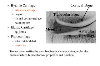
As a consequence of different functional requirements,:Three forms of cartilage have evolved.Each one exhibiting variation in matrix composition. In the matrix of hyaline cartilage, the most common form, type II collagen is the principal collagen type The more pliable and distensible elastic cartilage possesses, in addition to collagen type II, an abundance of elastic fiberswithin its matrix. Fibrocartilage, present in regions of the body subjected to pulling forces, is characterized by a matrix containing a dense network of coarse type I collagen fibers. Chapter 7 Cartilage Prof Abdel-Majeed Safer Histology & Hisctochemistry
Fig. 7-1 Chapter 7 Cartilage Prof Abdel-Majeed Safer Histology & Hisctochemistry
Fig. 7-1 Distribution of cartilage in adults. (a):three types of adult cartilage distributed in many areas of the skeleton, particularly in joints and where pliable support is useful, as in the ribs, ears, and nose. Cartilage support of other tissues throughout the respiratory system is also prominent. The photomicrographs show the main features of (b)hyaline cartilage, (c)fibrocartilage, and (d)elastic cartilage. Chapter 7 Cartilage Prof Abdel-Majeed Safer Histology & Hisctochemistry
avascular Cartilage is avascularand is nourished by the diffusion of nutrients from capillaries in adjacent connective tissue (perichondrium) or from synovial fluid in joint cavities.chondrocytesexhibit low metabolic activity. Cartilage also lacks lymphatic vessels and nerves. Chapter 7 Cartilage Prof Abdel-Majeed Safer Histology & Hisctochemistry
The perichondriumharborsthe vascular supply for the avascular cartilage and also contains nerves and lymphatic vessels. Articularcartilage, which covers the surfaces of the bones in movable joints, is devoid of perichondriumand is sustained by the diffusion of oxygen and nutrients from the synovial fluid. Chapter 7 Cartilage Prof Abdel-Majeed Safer Histology & Hisctochemistry
Fig. 7-2 Chapter 7 Cartilage Prof Abdel-Majeed Safer Histology & Hisctochemistry
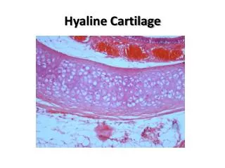
Hyaline CartilageHyaline cartilage is the most common and best studied .Fresh hyaline cartilage is bluish-white and translucent. In the embryo, it serves as a temporary skeleton until it is gradually replaced by bone.In adult mammals, hyaline cartilage is located in the articular surfaces of the movable joints, in the walls of larger respiratory passages (nose, larynx, trachea, bronchi), in the ventral ends of ribs, where they articulate with the sternum. in the epiphyseal plate, where it is responsible for the longitudinal growth of bone. Chapter 7 Cartilage Prof Abdel-Majeed Safer Histology & Hisctochemistry
Fig. 7-2 Chapter 7 Cartilage Prof Abdel-Majeed Safer Histology & Hisctochemistry
MatrixForty percent of the dry weight of hyaline cartilage consists of collagen embedded in a firm, hydrated gel of proteoglycansand structural glycoproteins. In routine histology preparations, the collagen is indiscernible for two reasons: the collagen is in the form of fibrils which have submicroscopic dimensions and the refractive index of the fibrils is almost the same as that of the surrounding substances. Type II collagen Hyaline cartilage contains primarily type II collagen, although small amounts of collagen types VI and IX are also present. Chapter 7 Cartilage Prof Abdel-Majeed Safer Histology & Hisctochemistry
Cartilage proteoglycans contain chondroitin 4-sulfate, chondroitin 6-sulfate, and keratan sulfate, covalently linked to core proteins. Proteoglycansare bound noncovalently to long molecules of hyaluronic acid by link proteins, forming very large proteoglycan aggregates such as aggrecan that interact with collagen. Structurally, proteoglycans resemble bottle brushes, the protein core being the stem and the radiating glycosaminoglycan (GAG) chains the bristles. Chapter 7 Cartilage Prof Abdel-Majeed Safer Histology & Hisctochemistry
Fig. 7-3 Chapter 7 Cartilage Prof Abdel-Majeed Safer Histology & Hisctochemistry
The high content of solvation water bound to the negative charges of the GAGs acts as a shock absorber or biomechanical spring; this is of great functional importance, especially in articularcartilages. Structural multiadhesive glycoprotein chondronectin:In addition to type II collagen and proteoglycan, an important component of cartilage matrix is the structural multiadhesive glycoprotein chondronectin. Like fibronectin in connective tissue, this macromolecule binds specifically to GAGs, collagen type II and integrins, mediating the adherence of chondrocytes to the ECM. solvation water Chapter 7 Cartilage Prof Abdel-Majeed Safer Histology & Hisctochemistry
Cartilage matrix is generally basophilic due to the high concentration of sulfatedGAGs and staining variations within the matrix reflect differences in the molecular composition. Immediately surrounding each chondrocyte the ECM is richer in GAGs and poor in collagen. These areas comprise the territorial matrix and usually stain differently from the rest of the matrix. Chapter 7 Cartilage Prof Abdel-Majeed Safer Histology & Hisctochemistry
Fig. 7-2 Chapter 7 Cartilage Prof Abdel-Majeed Safer Histology & Hisctochemistry
Cells:ChondrocytesAt the periphery of hyaline cartilage, young chondrocyteshave an elliptic shape, with the long axis parallel to the surface. Farther in, they are round and may appear in groups of up to eight cells originating from mitotic divisions of a single chondrocyte. These groups are called isogenous aggregates (Gr. isos, equal, + genos, family). Chondrocytessynthesize collagens and the other matrix molecules. Chapter 7 Cartilage Prof Abdel-Majeed Safer Histology & Hisctochemistry
As matrix is produced, cells in the aggregates are moved apart and occupy separate lacunae. Cartilage cells and the matrix often shrink during routine histologic preparation, resulting in both the irregular shape of the chondrocytes and their retraction from the matrix. Chapter 7 Cartilage Prof Abdel-Majeed Safer Histology & Hisctochemistry
Chondrocytesrespire under low oxygen tension. Hyaline cartilage cells metabolize glucose mainly by anaerobic glycolysis to produce lactic acid as the end product.Nutrients from the blood diffuse through the perichondrium to reach the more deeply placed cartilage cells. Transport of water and solutes is promoted by the pumping action of intermittent cartilage compression and decompression. Chapter 7 Cartilage Prof Abdel-Majeed Safer Histology & Hisctochemistry
Chondrocyte function is hormone dependent. Synthesis of sulfatedGAGs is accelerated by: growth hormone, thyroxin, testosterone slowed by: cortisone, hydrocortisone, estradiol. . Chapter 7 Cartilage Prof Abdel-Majeed Safer Histology & Hisctochemistry
Cartilage growth depends mainly on the pituitary-derived growth hormone somatotropin. This hormone does not act on cartilage cells directly but promotes the endocrine release in the liver of insulin-like growth factor-1 (IGF-1), sometimes called somatomedin C. IGF-1 acts directly on cartilage cells, promoting their growth Chapter 7 Cartilage Prof Abdel-Majeed Safer Histology & Hisctochemistry
PerichondriumExcept in the articular cartilage of joints, all hyaline cartilage is covered by a layer of dense connective tissue, the perichondrium, which is essential for the growth and maintenance of cartilage.It consists largely of collagen type I fibers and contains numerous fibroblasts. Although cells in the inner layer of the perichondrium resemble fibroblasts, they are precursors for chondroblasts which divide and differentiate into chondrocytes. Chapter 7 Cartilage Prof Abdel-Majeed Safer Histology & Hisctochemistry
Fig. 7-2 Chapter 7 Cartilage Prof Abdel-Majeed Safer Histology & Hisctochemistry
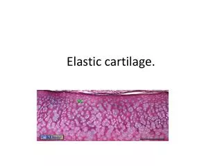
Elastic CartilageElastic cartilage is essentially very similar to hyaline cartilage except that it contains an abundant network of fine elastic fibersin addition to collagen type II fibrils Fresh elastic cartilage has a yellowish color owing to the presence ofelastinin the elastic fibers. Chapter 7 Cartilage Prof Abdel-Majeed Safer Histology & Hisctochemistry
Fig. 7-4 Chapter 7 Cartilage Prof Abdel-Majeed Safer Histology & Hisctochemistry
Elastic cartilage is frequently found to be gradually continuous with hyaline cartilage. Elastic cartilage possesses a perichondrium.Elastic cartilage is found in the auricle of the ear, the walls of the external auditory canals, the auditory (eustachian) tubes, the epiglottis, and the cuneiform cartilage in the larynx. Chapter 7 Cartilage Prof Abdel-Majeed Safer Histology & Hisctochemistry
FibrocartilageIntermediate between dense connective tissue and hyaline cartilage. It is found in intervertebral disks, in attachments of certain ligaments, and in the pubic symphysis. Fibrocartilageis always associated with dense connective tissue and the border between these two tissues is not clear-cut, showing a gradual transition. Chapter 7 Cartilage Prof Abdel-Majeed Safer Histology & Hisctochemistry
Chapter 7 Cartilage Prof Abdel-Majeed Safer Histology & Hisctochemistry
Fibrocartilage contains chondrocytes, either singly or in isogenous aggregates, usually arranged axially, in long rows separated by coarse collagen type I fibers and less proteoglycans than other forms of cartilage.Because it is richer in collagen type I, the fibrocartilage matrix is more acidophilic. Chapter 7 Cartilage Prof Abdel-Majeed Safer Histology & Hisctochemistry
Fig. 7-5 Chapter 7 Cartilage Prof Abdel-Majeed Safer Histology & Hisctochemistry
In fibrocartilage dense collagen fibers can form either irregular or parallel bundles between the axial aggregates of chondrocytes. The general orientation of the collagen depends on the stresses on fibrocartilage, since the collagen bundles take up a direction parallel to those stresses. There is no distinct perichondrium in fibrocartilage. Chapter 7 Cartilage Prof Abdel-Majeed Safer Histology & Hisctochemistry
Fig. 7-5 Chapter 7 Cartilage Prof Abdel-Majeed Safer Histology & Hisctochemistry
Intervertebral disks are composed of fibrocartilage primarily. Between the vertebrae and are held to them by ligaments. Each disk has two major histological components: the peripheral annulus fibrosus rich in bundles of type I collagen and the central nucleus pulposus with a gel-like matrix rich in hyaluronicacid.Intervertebraldisks act as lubricated cushions and shock absorbers preventing adjacent vertebrae from being damaged by abrasive forces or impact during movement of the spinal column. Chapter 7 Cartilage Prof Abdel-Majeed Safer Histology & Hisctochemistry
Cartilage Formation,Growth and RepairAll cartilage derives from the embryonic mesenchymein the process of chondrogenesis. The first indication of cell differentiation is the rounding up of the mesenchymal cells, which retract their extensions, multiply rapidly, and form cellular condensations. The cells formed by this direct differentiation of mesenchymal cells, now called chondroblasts, have a ribosome-rich basophilic cytoplasm. Synthesis and deposition of the matrix then begin to separate the chondroblasts from one another. Chapter 7 Cartilage Prof Abdel-Majeed Safer Histology & Hisctochemistry
Chapter 7 Cartilage Prof Abdel-Majeed Safer Histology & Hisctochemistry
Chapter 7 Cartilage Prof Abdel-Majeed Safer Histology & Hisctochemistry
During embryonic development, the differentiation of cartilage takes place primarily from the center outward; • therefore, the more central cells have the characteristics of chondrocytes, • whereas the peripheral cells are typical chondroblasts. • The superficial mesenchyme develops into the perichondrium. Chapter 7 Cartilage Prof Abdel-Majeed Safer Histology & Hisctochemistry
Fig. 7-6 Chapter 7 Cartilage Prof Abdel-Majeed Safer Histology & Hisctochemistry
Fig. 7-7 Chapter 7 Cartilage Prof Abdel-Majeed Safer Histology & Hisctochemistry
Further growth of cartilage is attributable to two processes: interstitial growth, resulting from the mitotic division of preexistingchondrocytes; appositional growth, resulting from the differentiation of perichondrial cells. In both cases, the synthesis of matrix contributes greatly to the growth of the cartilage. Interstitial growth is the less important of the two processes postnatally. It occurs during the early phases of cartilage formation, when it increases tissue mass by expanding the cartilage matrix from within Chapter 7 Cartilage Prof Abdel-Majeed Safer Histology & Hisctochemistry
Interstitial growth also occurs in the epiphyseal plates of long bones and within articular cartilage. In the epiphyseal plates, interstitial growth is important in increasing the length of long bones. In articular cartilage, as the cells and matrix near the articulating surface are gradually worn away, the cartilage must be replaced from within, since there is no perichondrium there to add cells by appositional growth. Chapter 7 Cartilage Prof Abdel-Majeed Safer Histology & Hisctochemistry
In cartilage found elsewhere in the body, interstitial growth becomes less pronounced, as the matrix becomes increasingly rigid from the cross-linking of matrix molecules. Cartilage then increases in girth only by appositional growth. Chondroblastsdifferentiate in the inner layers of the perichondrium, proliferate, and become chondrocytes once they have surrounded themselves with cartilaginous matrix and are incorporated into the existing cartilage., Chapter 7 Cartilage Prof Abdel-Majeed Safer Histology & Hisctochemistry
Fig. 7-2 Chapter 7 Cartilage Prof Abdel-Majeed Safer Histology & Hisctochemistry
regeneration Except in young children, damaged cartilage undergoes slow and often incomplete regeneration, by activity of cells in the perichondrium which invade the injured area and generate new cartilage. In extensively damaged areas—and occasionally in small areas—the perichondrium produces a scar of dense connective tissue instead of forming new cartilage. The poor regenerative capacity of cartilage is due in part to the avascularity of this tissue. Chapter 7 Cartilage Prof Abdel-Majeed Safer Histology & Hisctochemistry
End of chapter 7 Chapter 7 Cartilage Prof Abdel-Majeed Safer Histology & Hisctochemistry