Download
1 / 24
310 likes | 1.23k Views
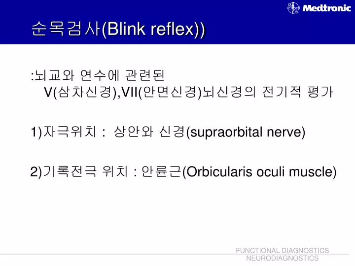
순목검사( Blink reflex)). :뇌교와 연수에 관련된 V( 삼차신경), VII( 안면신경)뇌신경의 전기적 평가 1)자극위치 : 상안와 신경( supraorbital nerve) 2) 기록전극 위치 : 안륜근( Orbicularis oculi muscle). 반복신경자극검사 ( Repetitive nerve stimulation test : Jolly test). :신경접합부의 이상을 알아보는 검사. 1) 저속반복신경자극 : 5 Hz 이하 자극,

E N D
순목검사(Blink reflex)) :뇌교와 연수에 관련된 V(삼차신경),VII(안면신경)뇌신경의 전기적 평가 1)자극위치 : 상안와 신경(supraorbital nerve) 2)기록전극 위치 : 안륜근(Orbicularis oculi muscle)
반복신경자극검사(Repetitive nerve stimulation test : Jolly test) :신경접합부의 이상을 알아보는 검사
1)저속반복신경자극 : 5Hz 이하 자극, 점감반응-중증근무력증 2)고속반복신경자극 : 5Hz이상자극, 점증반응 – 근무력 증후군, 보툴리누즘 중독
유발 전위 • 종 류 • 시각 유발전위(VEP) • 청각 유발전위(AEP) • 체성감각 유발전위(SEP) • 운동유발전위(MEP)
Averager (A.V.G.) Direct Signal Direct Signal AVG Group I AVG Group II AVG I + II Averaged Signal Amplified
VEP란? • 시각자극으로 인해 후두부에서 발생되는 전기적 변화를 기록한것 • 시기능을 객관적으로 측정
VEP 검사 방법 <전극부착법> • 활성전극 O1, Oz, O2, (LT, RT) • 기준전극 MF(mid-frontal) • 접지전극 Cz <자 극> • 자극 : 모니터를 이용한 시각자극 • 자극의 종류 1) 흑백의 격자무늬의 위상역전(pattern reversal, PR) 2) 섬광자극(Flash) 양측검사를 원칙으로 하며 검사실의 환경은 조용하고 어두워야 하며 환자의 얼굴근육의 이완으로 근 잡음을 방지해야 하고 시력이 좋지 않은 경우 시력을 교정한 상태에서 검사.
시각자극 ▼▼▼ 시신경-시신경교차-시각피질-전대뇌피질 ▼▼▼ N1(N75), P1(P100), N2(N140) ▼▼▼ 유발된 반응 관찰-임상에 활용
Goggles Flash Visual E.P. Optic Nerves & Stimulus Pattern: Checkers - Bares Optic Nerve Optic tract Lateral geniculate body Occipital lobe
Cz VER Right Eye O'z N145 O'1-Cz O1 Oz O2 A1 70 cm N75 O'z-Cz P100 Stim.: Checker Checker size: Visual angle * Stim. Freq.: Max. 2 Hz Dark room Averaging: 200 - 300 O'2-Cz Cz-A1 N100 100 200 ms Visual E.P. AVERAGING AVERAGING * See next slide
VEP Normal Values (P100) Full-field, screen size 9°, check size 26’ Mean Range SD P100 Latency ms 102.3 89-114 ±5.1 Latency diff. between two eyes 1.3 0-6 ±2.0 P100 Amplitude µV 10.1 3-21 ±4.2 Amplitude diff. 1.6 0-5.5 ±1.4
BAEP검사 방법 <전극부착법> 활성전극 A1(mastoid), A2(mastoid) 기준전극 Cz, 접지전극 Fpz <자 극> 주로 click sound (이어폰)로 1500번 자극 *자극편-hearing threshold + 65dB (똑똑…) *자극 반대편–자극귀의 dB에서 –40dB의 white noise(치치..) <유발 파형> I, II, III, IV, V 중 절대잠복기인 I, III, V 파간 잠복기인 I-III, III-V, I-V이 중요함.
청각자극 ▼▼▼ 청각신경-뇌간부 ▼▼▼ I, III, V / I-III, III-V, I-V ▼▼▼ 유발된 반응 관찰-임상에 활용
BAEP(뇌간청각 유발전위) 자극편(click sound) 자극반대편(white noise)
Wave I : 청각신경(cochlear nerve) 원위부 Wave III : 교 (pons) Wave V : 중 뇌 (midbrain)
SEP (체성감각유발전위) • SEP 또는 SSEP. • 전기자극 – 말초신경 – 척수 - 대뇌피질로 전달, 중추신경계와 말초신경계의 기능을 모두 대변. • 상지 SEP arm • 하지 SEP leg • 양측검사를 기본으로 한다.
SEP in Upper extremity (참고서적 Spehlmann’s Evoked potential primer) • SEP 상지 (활성-기준) 우측 CH 1 : C3’ (C3 2cm 후방)– A1(Lt. mastoid) : sensory cortex Ch 2 : C5 – Fpz : cervical cord Ch 3 : EP2 – EP1 : peripheral nerve 접지 : Cz 자극 : 손목의 정중신경(median nerve) • SEP 상지 (활성-기준) 좌측 CH 1 : C4’ (C4 2cm 후방) – A2 (Rt. mastoid) : sensory cortex Ch 2 : C5 – Fpz : cervical cord Ch 3 : EP1 – EP2: peripheral nerve 접지 : Cz 자극 : 손목의 정중신경(median nerve)
SEP in Lower extremity • SEP 하지 (활성-기준) 우측 CH 1 : Cz’ (Cz 2cm 후방) – Fpz : senosry cortex Ch 2 : T12 – Rt. iliac crest : peripheral nerve-spinal cord 접지 : Rt. knee 자극 : 발목 내측의 후경골신경(poterior tibial nerve) • SEP 상지 (활성-기준) 좌측 CH 1 : Cz’ (Cz 2cm 후방) – Fpz : senosry cortex Ch 2 : T12 – Lt. iliac crest : peripheral nerve-spinal cord 접지 : Lt. knee 자극 : 발목 내측의 후경골신경(poterior tibial nerve)
N Tibial Nerve 7 cm Fpz L R C5S CPi-Fpz Fz 5 cm Cz P37 2 cm CPi CPz CPc T12S CPz-Fpz I P37 N34 IC P31 Fpz-C5S Stim. Freq.: 3 Hz Intensity: Threshold Averaging: 200 - 500 AVG Sens.: 2 - 5 µV/D LP T12S-IC 20 40 60 ms Stim. Somatosensory E.P. Lower Extremities AVERAGING AVERAGING
N Median Nerve 7 cm N19 Fpz C5S L R CPc-CPi CPi-Epc C5S-Epc Epi-Epc Fz Epi Epc 5 cm C3 C4 2 cm N18 CPi CPc I P14 Stim. N13 EP Stim. Freq.: 3 Hz Intensity: Threshold Averaging: 200 - 500 AVG Sens.: 2 - 5 µV/D 10 20 30 ms Somatosensory E.P. Upper Extremities
SEP Normal Values Latency msAmplitude µV Mean SD Range Mean SD Range Median Nerve EP 9.6 ±0.7 5.4 ±2.5 N13 13.2 ±0.8 2.9 ±1.3 N19 18.9±1.0 2.8 ±1.6 Tibial Nerve LP 10.8 ±0.9 8.6-13.1 N34 33.5 ±1.5 30.3-41.3 P37 36.3±2.4 30.5-41.7