Download
1 / 28
280 likes | 381 Views
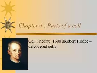
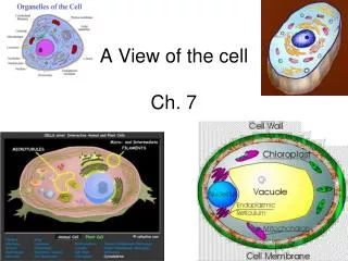
Chapter 4- A View of the Cell. 4.1-The Discovery of Cells. Cells are the basic units of living organisms Development of the microscope allowed scientists to view cells. Cell Theory. 1665-Robert Hooke Uses compound microscope to observe cork Hollow boxes…..CELLS!!

E N D
4.1-The Discovery of Cells • Cells are the basic units of living organisms • Development of the microscope allowed scientists to view cells
Cell Theory • 1665-Robert Hooke • Uses compound microscope to observe cork • Hollow boxes…..CELLS!! • 1830s-Schleiden and Schwann • Schleiden-all plants are composed of cells! • Schwann-all animals are composed of cells!
Three Parts of Cell Theory • 1. All organisms composed of one or more cells. • 2. Cell is the basic unit of organization of organisms. • 3. All cells come from preexisting cells.
History of the Microscope • 1665-Hooke’s Microscope • Three lenses • Poor Quality • Little detail
1700-Anton van Leeuwenhoek • Built over 240 microscopes! • Single Lens • Better Quality • Observed red blood cells & bacteria • Mid 1800s-Compound Light Microscope • Series of lenses • Light passes through object then lens • More detail
1940s-Electron Microscopes • Beam of electrons through magnetic field • Specimen in vacuum • Only view dead cells or organisms • 1.SEM-Scanning Electron Microscope • Surface of specimen-3D Picture! • 2.TEM-Transmission Electron Microscope • Through the specimen • Magnify 100s or 1,000s of times! • 3.STM-Scanning Tunneling Microscope • Arrangement of atoms on surface • Map hills and valleys
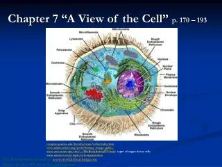
Basic Cell Types… • Look at internal organization! • Prokaryotes-lacks internal structures • Eukaryotes-membrane bound, internal structures. • Structures called organelles • Largest organelle = NUCLEUS!
Section 4.2-Eukaryotic Cell Structure • Must have boundaries! • Plasma Membrane • Boundary between cell & environment • Flexibility • Controls movement of materials • Cell Wall-only plant cells! • Rigid, surrounds membrane • Thicker, stronger network for structural support
GET UNDER CONTROL!!!! • Nucleus-contains cell’s DNA, manages cell function • Surrounded by nuclear envelope • Double membrane-pores allow movement • Chromatin-long strands of DNA • Packed into chromosomes
Nucleolus • Region inside nucleus • Produces particles for protein synthesis • Particles are called ribosomes • Cell assembles enzymes according to DNA • Not bound by membrane
Assembly, Transport, & Storage • Cytoplasm-outside nucleus, surrounds organelles • Clear liquid • Important chemical reactions (protein assembly) take place here • Endoplasmic Reticulum (ER)- • Network of interconnected compartments • Surface folded into cell • Tissues in a box! • Site for lipid synthesis in cell
Smooth or Rough? • Smooth ER • Lacks ribosomes on surface • Rough ER • Studded with ribosomes on surface • ER acts as cell’s delivery system!
Structures for Storage • Golgi Apparatus • Closely stacked, flattened membrane sacs • Receives proteins and lipids • Modifies them chemically, repackages • Distributes them throughout membrane • Vacuole-sac of fluid • Temporary storage of food, enzymes etc. • Animal cell-small and numerous • Plant cell-single large vacuole for water
Golgi Apparatus Vacuole
Reduce, Reuse, RECYCLE! • Lysosomes • Digest excess or worn out cell parts, food particles, and invading viruses/bacteria • Membrane protects digestive enzymes from rest of cell
Energy Transformers • Mitochondria • Break down food-convert to energy! • Peanut-shaped • Highly folded inner membrane for more energy storage • Chloroplasts-plants only! • Transform light energy into chemical energy • Chlorophyll-traps energy from sun • Green color!
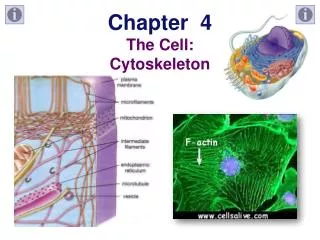
Support and Locomotion • Cytoplasm • 1. Cytoskeleton-network of fibrous elements • Act as scaffold for organelle support • 2. Microtubules-thin, hollow cylinders • Made of protein • 3. Microfilaments-thin, solid fibers • Both make up most of cytoskeleton
LET’S GET MOVING! • Cilia • Short, numerous hair-like projections • On cell’s surface • Movement like “the wave!” • Flagella • Longer projections • Whip-like motion • One or two per cell
Cellular Organization • 1. Single-celled organisms: Unicellular • 2. Many-celled organisms: Multicellular • 3. Cells functioning together for activity: Tissues • 4. Two or more tissues functioning together: Organs • 5. Organs working together to carry out major life functions:Organ System