Download
1 / 53
530 likes | 648 Views
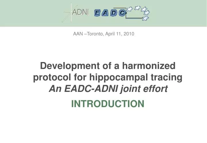
AAN –Toronto, April 11, 2010. Development of a harmonized protocol for hippocampal tracing An EADC-ADNI joint effort INTRODUCTION. BACKGROUND Hippocampal volume as a diagnostic marker. Structure: MR hippo volumetry. Function: FDG PET. Amyloid deposits: PET. Biochemistry: CSF

E N D
AAN –Toronto, April 11, 2010 Development of a harmonized protocol for hippocampal tracing An EADC-ADNI joint effortINTRODUCTION
BACKGROUND Hippocampal volume as a diagnostic marker Structure: MR hippo volumetry Function: FDG PET Amyloid deposits: PET Biochemistry: CSF tau/Abeta Dubois, Feldman et al., Lancet Neurol 2007
BACKGROUND The effect of segmentation protocols on hippocampal volume Geuze et al., Mol Psychiatry 2005;10:147-59
BACKGROUND Hippocampal volume as an outcome measure in trials of disease modifiers * CHV= Change in hippocampal volume; § referenced link not working
BACKGROUND Hippocampal volume (manual segmentation) as gold standard for automated segmentation algorithms Morey et al., NeuroImage 2009
AIMS To Harmonize Available Protocols for Manual Hippocampal Segmentation To Test the Validity of the Harmonized Protocol
THE WORKGROUP EADC F Barkhof/P Scheltens Amsterdam B Dubois/S Leherici Paris N Fox London GB Frisoni Brescia H Hampel/J Pantel Frankfurt A Simmons London H Soininen Kuopio S Teipel Rostock LO Wahlund Stockholm
THE WORKGROUP ADNI CR Jack Jr Rochester, MN G Barzokis UCLA, CA D Bennet Chicago, IL J Csernansky NorthW U, WA C DeCarli UC Davis, CA M De Leon New York, NY J Kaye Portland, OR R Killiany Boston USM, MA PM Thompson LoNI, UCLA, CA M Weiner/S Mueller UCSF/VAMC
THE WORKGROUP other relevant hippo experts R Camicioli Univ Alberta, Canada J O’Brien NewCastle, UK J Pruessner McGill, QC, Canada
THE WORKGROUP Advisory Boards EADC-ADNI P.I.s B Vellas Toulouse, France B Winblad Stockholm, Sweden M Weiner UCSF, US Population-based studies M Breteler / T Den Heijer Rotterdam Scan Study P Sachdev / JJ Maller PATH through life L Launer NIA Bethesda W Jagust Berkeley
THE WORKGROUP Boards Statistical Board S Duchesne Laval Univ, Canada L Collins MNI, McGill, Montreal P Pasqualetti AFaR, Rome Clinical Advisor PJ Visser Maastricht, The Netherlands Dissemination/Education G Waldemar Copenhagen, Denmark
THE WORKGROUP Personnel in Brescia Marina Boccardi psychologist, PhD Rossana Ganzola psychologist Enrica Cavedo psychologist Michela Pievani mathematician Anna Caroli mathematician Alberto Redolfi bioinformaticist
AAN –Toronto, April 11, 2010 Development of a harmonized protocol for hippocampal tracing An EADC-ADNI joint effortMETHODS
METHODS Operationalization of protocols’ differences and extraction of tracing units Quantification of tracing units’ features Harmonized protocol
OPERATIONALIZATION Selection of segmentation protocols • Selection criteria • Being based on 3D T1 MRI with field strength greater than 1 Tesla • Providing explicit description of landmarks for tracing of hippocampal borders • Being validated on AD/MCI samples, or being widely adopted in the literature about AD.
OPERATIONALIZATION Protocols’ features extraction Plane of tracing: AC-PC line; Normalization to the Talairach space Tracing start from the tail of hippocampus to the hippocampal head In the following slices, you can find the hippocampal tracing on consecutive coronal slices (1.5T) of the control subject and AD patient according to Pruessner's criteria
OPERATIONALIZATION Prototypical tracings Tracings carried out on a control and an AD subject from the ADNI dataset Tracings sent to the Authors for correction or approval
CTRL Most posterior slice : slice where an ovoid mass of gray matter (1) started to appear inferomedially of the atrium of lateral ventricle (2) 2= Atrium of lateral ventricle 4= Parahippocampal gyrus 5= Gyrus dentatus 6= Cornu Ammonis 7= Gyrus fasciolaris 8= Gyrus of Andreas-Retzius 9= Isthmus The fornix can be excluded by a horizontal line (in fuchsia) from the superior border of the quadrigeminal cistern (3) to the lateral ventricle (2). Part of gyrus fasciolaris (7) and of Andreas-Retzius gyrus (8) can be excluded by a vertical line (in red) from the medial end of the lateral ventricle (2) down to the parahippocampal gyrus (4). Sagittal view
AD Most posterior slice : slice where an ovoid mass of gray matter (1) started to appear inferomedially of the atrium of lateral ventricle (2) 2= Atrium of lateral ventricle 4= Parahippocampal gyrus 5= Gyrus dentatus 6= Cornu Ammonis 7= Gyrus fasciolaris 8= Gyrus of Andreas Retzius 9= Isthmus In AD subjects the cerebral structures are atrophic, lateral ventricle (2) and ambient cistern (3) result enlarged. Therefore, the lines used to exclude the gyrus fasciolaris, the Andreas- Retzius gyrus and the fornix do not intersect the hippocampal tail. That being so, is there another way to exclude the gyrus fasciolaris, the Andreas-Retzius gyrus and the fornix in these subjects? Sagittal view
OPERATIONALIZATION Authors’ check For each protocol: features extraction, tracing and author’s certification CTRL X 10 AD
OPERATIONALIZATION Extraction of similarities and differences Harmonized language Reduction of redundancy Tracing units and subunits
OPERATIONALIZATION Extraction of similarities and differences
METHODS Operationalization of protocols’ differences Quantification of tracing units’ features Harmonized protocol
QUANTIFICATION OF TRACING UNITS’ FEATURES Contribution to hippo volume in control and AD Effect on re-test variability Contribution to differences between AD and controls
METHODS Operationalization of protocols’ differences Quantification of tracing units’ features Harmonized protocol
HARMONIZED PROTOCOL Participants to a Delphi panel based on: expertise in hippocampal anatomy manual tracing scientific production Assisted decisions based on: Low variability, highly informative.. ..included in harmonized protocol High variability, little informative.. ..excluded from harmonized prot
HARMONIZED PROTOCOL Validation & implementation Validation with neuropathological data Comparison with currently used protocols Public tracings and probability maps Standard environment for tracing, learning, and certification
AAN –Toronto, April 11, 2010 Development of a harmonized protocol for hippocampal tracing An EADC-ADNI joint effortEARLY RESULTS
EARLY RESULTS Protocols’ features extraction & differences operationalization Modeling & computations of the operationalized differences Discussion
PROTOCOLS’ FEATURES EXTRACTION B Bartzokis G, et al. Psychiatry Res 1998;82:11-24 C Convit A, et al. Neurobiol Aging 1997;18:131-8 H Haller JW, et al. Radiology 1997;202:504-10 J Jack CR Jr. Epilepsia 1994;35 - Suppl 6:S21-9 K Killiany RJ, et al. Arch Neurol 1993;50:949-54 L Lehéricy S, et al. Am J Neuroradiol 1994;15:929-37 M Malykhin NV, et al. Psychiatry Res 2007;155:155-65 Pa Pantel J, et al. Hippocampus 2000;10:752-8 Pr Pruessner JC, et al. Cereb Cortex 2000;10:433-42 S Soininen HS, et al. Neurology 1994;44:1660-8
PROTOCOLS’ FEATURES EXTRACTION http://www.hippocampal-protocol.net
PROTOCOLS’ FEATURES EXTRACTION Harmonized language Reduction of redundancy Tracing units and subunits
PROTOCOLS’ FEATURES EXTRACTION Most anterior slice No longer an issue thanks to software for 3D navigation
PROTOCOLS’ FEATURES EXTRACTION Plane of tracing Axis of hippocampus AC-PC B. Bartzokis et al 1998 C. Convit et al 1997 J. Jack 1994 L. Lehéricy et al 1994 S. Soininen H et al 1994 H. Haller-Csernansky et al 1997 K. Killiany et al 1993 M. Malykhin et al2007 Pa. Pantel et al2000 Pr. Pruessner et al2000
PROTOCOLS’ FEATURES EXTRACTION Most posterior slice
A. Level where the inferior (1) and superior (2) colliculi are jointly visualized [B] B. Slice where the crus of fornix is visible in full profile (3) [C, K, L] C. Slice where the crura of fornices on both sides are seen in full profile (4, 5) [J, S] D. Slice where an ovoid mass of gray matter (6) appears inferomedially to the trigone of the lateral ventricle (posterior to anterior) [H, M, Pa, Pr]
PROTOCOLS’ FEATURES EXTRACTION Alveus Yellow = Alveus
PROTOCOLS’ FEATURES EXTRACTION Fimbria Yellow = Fimbria
PROTOCOLS’ FEATURES EXTRACTION Upper border
PROTOCOLS’ FEATURES EXTRACTION Medial border Vertical line [C] 45° line [Pr] Line with same inclination WM [K, L, M] Horizontal line [B, H, J] Morphological details [Pa, S]
EARLY RESULTS Protocols’ features extraction & differences operationalization Modeling & computations of the operationalized differences Discussion
Modeling by Simon Duchesne and Nicolas RobitailleUniversité Laval and Centre de Recherche Université Laval – Robert GiffardQuebec City, Canada
MODELING & COMPUTATIONS B C K L S J Pr M Pa H