Download
1 / 26
450 likes | 1.72k Views
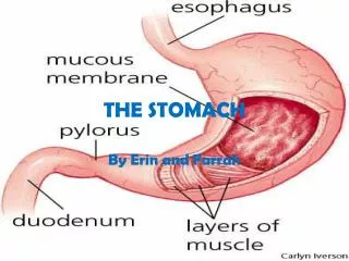
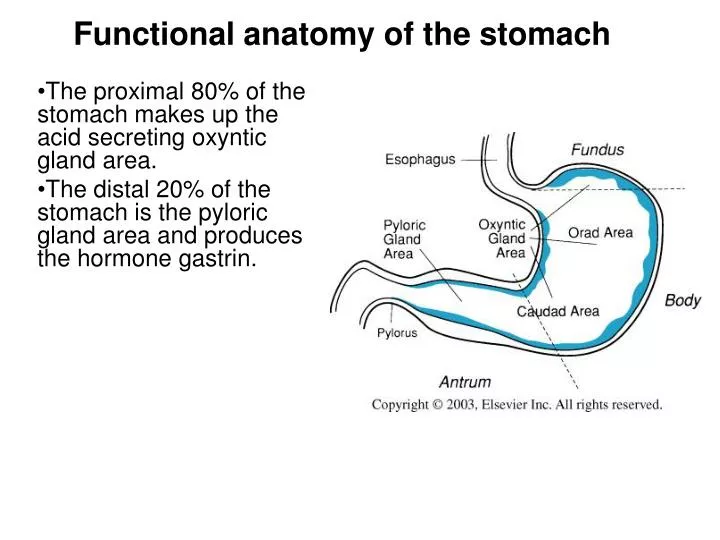
Functional anatomy of the stomach. The proximal 80% of the stomach makes up the acid secreting oxyntic gland area. The distal 20% of the stomach is the pyloric gland area and produces the hormone gastrin. Gastric Secretion and Function .

E N D
Functional anatomy of the stomach The proximal 80% of the stomach makes up the acid secreting oxyntic gland area. The distal 20% of the stomach is the pyloric gland area and produces the hormone gastrin.
Gastric Secretion and Function Body (Corpus) has oxyntic glands which contain parietal cells that secrete acid and intrinsic factor, mucus cells, Entero-chromaffin like (ECL) cells (histamine) and chief cells (pepsinogen) Fundus function is storage Antrum has pyloric glands containing mucus cells (mucus and pepsinogen), G cells (gastrin), and D cells (somatostatin), but no acid secreting parietal cells Pylorus controlled to release chyme slowly enough for digestion to occur.
Gastric Secretion and Function Components of gastric juice HCl activates pepsinogen, kills bacteria, and solubilizes and digests some food. Pepsin begins the digestion of protein. Mucus protects the lining of the stomach. Intrinsic factor is necessary for the absorption of vitamin B12.
Gastric “Oxyntic” gland • Surface epithelial cells secrete visible mucus. • Mucous neck cells secrete soluble mucus, and this region contains the proliferative cells as well as a stem cell. • The parietal cells secrete 1 to 2 liters of 150 to 160 mmol per liter of hydrochloric acid per day as well as intrinsic factor. • Peptic or chief cells secrete pepsinogen. • ECL cells secrete histamine
Parietal Cells • The parietal cell has a specialized tubulovesicular structure that increases apical membrane surface area 6-10 fold when the cell is stimulated to secrete acid. • H-K ATPase (proton pump) and Cl- channels are inserted into membrane for acid secretion.
Parietal Cells • Parietal cells also secret intrinsic factor, a 55 kD mucoprotein which complexes with vitamin B12 and is required for B12 absorption in the ileum. • Intrinsic factor secretion is the only stomach function necessary for survival!
Composition of gastric juice • Basal acid secretion is about 2-3 mEq/h but increases 10-100 times when stimulated. • Increasing rates of secretion of gastric juice cause the H+ concentration to rise and the Na+ concentration to fall. • Prolonged vomiting causes hypokalemia due to high K+
Mechanism of Acid Secretion • An apical H-K pump (ATPase) is responsible for gastric acid secretion by parietal cells. • Blocked by omeprazole, a proton pump inhibitor. • Secretion is carbonic anhydrase dependent. • A small non-parietal secretion at the base of the glands has an ion concentration near plasma • 150 mM HCl and 10-20 mM KCl are added depending on stimulation
Mechanism of acid secretion • H+ is extrude against a strong concentration gradient in exchange for K+ which diffuses back to lumen. • H+ is derived from H2CO3 dissociation catalyzed by carbonic anhydrase after CO2 diffuses into cell • Pure parietal cell secretion has 150 mEq/l H+ vs 0.00004 mEq/l in plasma, or 0.17 N HCl. • Water follows ion secretion into lumen
Mechanism of acid secretion • HCO3- extruded from cell base is exchanged for Cl- which diffuses into lumen through Cl- channels. • HCO3- entering the blood is responsible for the “postprandialalkaline tide“ after eating and a negative gastric arterial-venous CO2 gradient.
Secretagogues • Acetylcholine, histamine and gastrin directly and indirectly induce acid secretion by parietal cells. • Acetylcholine acts through the M3 receptor • Gastrin acts through the CKKB receptor • Both are Gaq linked to increases in Ca2+ and diacylglycerol through the PLC – IP3 pathway • Prostaglandin E2 acts on Gi to inhibit AC and acid secretion.
Secretagogues • Histamine acts through a Gas and increases cAMP and has most powerful overall action. • Gastrin is released by both antral and duodenal G cells. • Histamine is released by enterochromaffin-like cells (ECL) in the corpus which are also activated by acetylcholine (vagus n.) and gastrin. • Cimetidine, an H2 receptor blocker reduces acid secretion
Secretagogues • Gastrin and cholecystokinin act on CCKB receptors. • Gastrin is secreted by G cells in the antrum and duodenum and in response to gastrin-releasing peptide (GRP) from nerve fibers. • G17, G34 and CCK have same terminal sequence for CCKB and CCKA receptor binding but gastrin has orders of magnitude greater effect than CCK for acid secretion
Summary of control of acid secretion • Stomach distention via vagovagal reflex increases parietal cell acid production by: • direct stimulation of ECL cell to release histamine; • direct stimulation of G cells to release gastrin via GRP; • Inhibition of somatostatin release from D cells in corpus and antrum. • Antral exposure to peptides from protein digestion stimulates G cells to release gastrin and increases acid secretion by direct stimulation of G cells and stimulation of ECL cells to secrete histamine. • High acid feeds back to stimulate D cells to release somatostatin and inhibits gastrin release to limit acid secretion.
Gastric secretion in response to a meal • Food in the stomach buffers acid and raises the pH • This suppresses somatostatin production from inhibitor somatostatin (D) cells causing release of gastrin and acid production
Gastric secretion in response to a meal • Stomach distention, and digestion products trigger a tremendous oupouring of secretion volume and acid. • As food leaves stomach and acid increases, pH falls, stimulates D cells and acid production is inhibited by somatostatin. Stomach distention also decreases.
Gastric Emptying • Fats and to a lesser extent protein digestion are rate limiting and the major controllers of emptying. • Isotonic solutions pass rapidly through the stomach. • CHO is rapidly digested with as much as 50% being digested in the stomach by salivary amylase before the stomach contents become acidified. • Elevated CCK stimulated by FFA constricts the pylorus and limits emptying • GIP and peptide YY from the intestine are also simulated by FFA and inhibit emptying.
Gastric Emptying • Secretin inhibits acid secretion and emptying if the duodenum is too acid. • Osmoreceptors in the duodenum initiate a reflex inhibition of emptying if the chyme is hypertonic by unknown enterogastone. • Gastrin released by amino acids increases pyloric tone • Vagal stimulation increases pyloric tone.
Pepsinogen secretion • Inactive pepsinogen is stored and secreted by chief cells and mucus cells • Pepsinogen protects the chief cell from digestion. • At a low pH (3.0-5.0) pepsinogen is split to active pepsin to further split pepsinogen. • Pepsin activated at pH <3.5, inactive at pH 7.0 • Acetylcholine via vagal stimulation is the strongest stimulus for pepsin secretion, but secretin also stimulates pepsin secretion.
Pepsinogen secretion • Acid also stimulates pepsin secretion by a local cholinergic vago-vagal reflex and by stimulating secretin release from S cells in the duodenum • Second messengers are cAMP (secretin) and Ca2+ (acetylcholine) for secretion of multiple pepsinogens • Salivary amylase initiates starch digestion, and lingual and gastric lipases also digest lipids in the stomach
Protection of the gastric surface epithelium • Acid is squirted through mucus layer as a jet • A mucus gel layer 50-200 mm thick creates a diffusion barrier for H+ and a pH gradient of 1 to 7.2 • Impermeable apical cell membranes and tight junctions limit H+ ion diffusion across the epithelium • Vagal stimulation and irritation stimulate gastric mucous cells to secrete mucin, a glycoprotein that is part of the mucosal barrier
Protection of the gastric surface epithelium • HCO3- accumulates near the cell surface • Gastric surface epithelial cells secrete HCO3- when stimulated by acetylcholine, acids, and prostaglandins • Mucus protects the gastric surface epithelium by trapping an HCO3- rich fluid near the apical border of these epithelial cells
Peptic ulcer disease- breakdown of barrier by acid, stress, alcohol or aspirin can cause ulcers in the stomach and duodenum. • Most stomach ulcers are now known to be due to infection with Helicobacter pylori and can be treated with antibiotics, proton pump inhibitors and H2 blockers. • Zollinger-Ellison syndrome is a condition of excess stomach acid and ulcers due to gastrin producing tumors. • Pernicious anemia is a macrocytic anemia due to lack of parietal cells (intrinsic factor) and loss of B12 absorption.
SUMMARY •Gastric acid is secreted in concentrations as high as 150 mmol/L by the parietal cells, which contain a proton pump (H+,K+-ATPase) in their apical membranes. Acid converts inactive pepsinogen to pepsin, solubilizes some food, and kills bacteria. •The major stimulants of acid secretion are the hormone gastrin, the vagal mediator acetylcholine, and histamine, which is released from the enterochromaffin-like (ECL) cells by gastrin and acetylcholine and acts as a paracrine to stimulate the parietal cells. • The cephalic phase of gastric secretion is mediated by the vagus nerve, which stimulates the parietal cell via acetylcholine and releases gastrin from the G cells via gastrin-releasing peptide (GRP).