Download
1 / 22
220 likes | 915 Views
The Acute Abdomen. Major causes of the 'acute abdomen'. Acute cholecystitis Acute appendicitis or Meckel's diverticulitis Acute pancreatitis Peptic ulcer disease Pelvic inflammatory disease Intestinal obstruction , including paralytic ileus (adynamic obstruction)

E N D
Major causes of the 'acute abdomen' • Acute cholecystitis • Acute appendicitis or Meckel's diverticulitis • Acute pancreatitis • Peptic ulcer disease • Pelvic inflammatory disease • Intestinal obstruction, including paralytic ileus (adynamic obstruction) • Acute intestinal ischaemia/infarction or vasculitis • Gastrointestinal haemorrhage • Non-surgical disease, e.g. myocardial infarction, pericarditis, pneumonia, sickle cell crisis, hepatitis, inflammatory bowel disease, opiate withdrawal, typhoid, acute intermittent porphyria, HIV-associated lymphadenopathy or enteritis
Gallstones and Cholecystitis • Gallstones may cause no symptoms and are occasionally discovered • The second commonest presentation is acute cholecystitis, caused by distension of the gallbladder with subsequent necrosis and ischaemia of the mucosal wall.
Biliary colic • The pain starts suddenly in the epigastrium, • Vomiting often accompanies the pain,
Investigations • Urinalysis, chest X-ray and ECG may help exclude other diseases. • Ultrasound is the best way to demonstrate stones. • Ultrasonography can also allow measurement of the diameter of the common bile duct
Investigations • Endoscopic retrograde cholangiopancreatography (ERCP) is currently the only reliable and widely available investigation for duct stones. • CT may be useful when filling the bile duct is unsuccessful • Oral cholecystograms (contrast given orally is concentrated in a healthy GB
Cholecystitis • The main difference from biliary colic is the inflammatory component • If the stone moves to the common bile duct (CBD) jaundice may occur • Ultrasonography confirms dilatation of the common bile duct (>7mm diameter )
Non-surgical • Biliary colic and acute cholecystitis - these conditions will usually respond to an opioid such as morphine • Pain continuing for over 24 hours or accompanied by fever usually necessitates hospital admission. • Require antibiotics
Surgical • Laparoscopic cholecystectomy • Early surgery (within seven days of the onset of symptoms) appears to be safe and shortens hospital stay, but further studies are needed. • Postoperative complications are rare but do occur. The most significant is injury to the bile duct which occurs at a rate of 0.2% in both open and laparoscopic surgery.
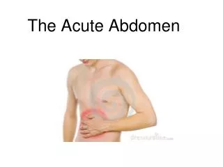
Acute Appendicitis • Sudden inflammation of the appendix usually caused by obstruction of the lumen resulting in invasion of the appendix wall by the gut flora. • Appendicitis is more common in men. • Appendicectomy is performed more often in women
Classic symptoms • Pain: • Early peri-umbilical pain moves after hours or sometimes days to the right iliac fossa • Vomiting, anorexia • Temperature and pulse are normal at first • Rectal examination: • TR-Appendicectomy
Intestinal obstruction and ileus • Obstruction to free passage of contents can occur at any level of the gut but problems such as oesophageal stricture, oesophageal carcinoma, carcinoma of stomach and pyloric stenosis will not be considered here but only obstruction beyond the duodenum. Ileus means intestinal blockage. The term paralytic ileus is used when the problem is inactivity of the bowel. Ileus is often used as a synonym. This may also be called intestinal pseudo-obstruction.
Risk factors: • Small intestinal obstruction is caused by adhesions in 60%, strangulated hernia in 20%, malignancy in 5% and volvulus in 5%. • Large intestinal obstruction is most often the result of colo-rectal malignancies • Sigmoid and caecal volvulus describes rotation of the gut on its mesenteric axis
Paralytic ileus • Paralytic ileus describes the condition in which the bowel ceases to function and there is no peristalsis. • chest infection • acute myocardial infarction • Stroke, trauma • acute renal failure • severe hypothyroidism • electrolyte disturbance • diabetic ketoacidosis
Clinical • Central abdominal pain • Vomiting tends to be early in high level obstruction • Abdominal distension • Check hernial orifices • Plain abdominal x-ray is a very important investigation
Management: • Resuscitation is very important • Nasogastric tube will reduce vomiting • Early surgery is required if there is local or generalised peritonitis, • The management of patients with obstruction due to malignancy who are unfit for surgery
Prognosis: • The prognosis of advanced carcinoma of the colon remains poor. 25% have distant metastases • 50% of sigmoid volvulus will recur in the next 2 years • 60% of stomas are never reversed • Older patients are less able to withstand the rigours of serious illness and major surgery
Acute Pancreatitis • Pathogenesis -Gallbladder disease and excess alcohol consumption account for most cases and typically cause periductal necrosis. Biliary disease and alcohol abuse together account for 70%-80% of cases,
Signs • Take temperature to exclude hypothermia • Probable tachycardia • Epigastric or generalised abdominal tenderness, often with rigidity. • In severe cases: gross hypotension, pyrexia, tachypnoea • Hypoxaemia is characteristic of acute pancreatitis
Investigations • Serum amylase >4 x normal: • Raised bilirubin and/or serum aminotransferase suggest gall stones. • Hypocalcaemia is relatively common. • CT with contrast enhancement may be diagnostic where clinical and biochemical results are equivocal on admission
Management • Pain relief with pethidine or buprenorphine ± IV benzodiazepines. Morphine relatively contraindicated because of possible spastic effect on sphincter of Oddi. • Nasogastric tube only for severe vomiting. • Antibiotics for specific infections.
Complications • Pancreatic necrosis • Infected necrosis • Pancreatic abscess • Acute pseudocyst • Pancreatic ascites • Systemic complications Respiratory, Cardiovascular, Disseminated intravascular coagulopathy (DIC), Hypocalcaemia