Download
1 / 45
500 likes | 743 Views
ST Segment Elevations in ECG. San Juan County EMS Paramedic Run Review 2011. Introduction. ST segment of the cardiac cycle represents the period between depolarization and repolarization of the left ventricle In normal state, ST segment is isoelectric relative to PR segment. Introduction.

E N D
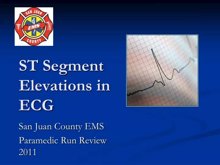
ST Segment Elevations in ECG San Juan County EMS Paramedic Run Review 2011
Introduction • ST segment of the cardiac cycle represents the period between depolarization and repolarization of the left ventricle • In normal state, ST segment is isoelectric relative to PR segment
Introduction • Most ST segment elevation is a result of non-AMI causes • Otto LA, Aufderheide TP. Evaluation of ST segment elevation criteria for the prehospital electrocardiographic diagnosis fo acute myocardial infarction. Ann Emerg Med 1994; 23 (1):17-24. • Chan TC, Brady WJ, Harrigan RA et al. ECG in Emergency Medicine and Acute Care. 1st ed. Pennsylvania: Elsevier Mosby; 2005.
Introduction • Of 123 adult chest pain patientswithST segment elevation ≥ 1mm, 63 patients (51%) did not have myocardial infarctions. • These non-MI were mainly • LBBB (21%) and • LVH (33%). • Otto LA, Aufderheide TP. Evaluation of ST segment elevation criteria for the prehospital electrocardiographic diagnosis fo acute myocardial infarction. Ann Emerg Med 1994; 23 (1):17-24.
Acute Pericarditis Benign Early Repolarization Left Bundle Branch Block with AMI (Sgarbossa et al’s criteria) Left Ventricular Hypertrophy Left Ventricular Aneurysm Brugada Syndrome Hyperkalemia Hypothermia CNS pathologies Prinzmetal Angina Post electrical cardioversion Causes of ST Segment Elevation
Acute Myocardial Infarction • Initial ST elevation as part of the classic evolutionary pattern of acute myocardial infarction was first described by Pardee in 1920 • Pardee HEB. An electrocardiographic sign of coronary artery obstruction. Arch Intern Med 1920; 26: 244–57.
Acute Myocardial Infarction • The exact reasons AMI produces ST segment elevation are complex and not fully understood • MI alters the electrical charge on the myocardial cell membranes and produce an abnormal current flow • Goldberger: Clinical Electrocardiography: A Simplified Approach, 6th edition, 1999.
TP segment or PR segment? • ST segment elevation measured: • At J point – if relative to PR segment • At 0.06 – 0.08s from J point – if relative to TP segment • Chan TC, Brady WJ, Harrigan RA et al. ECG in Emergency Medicine and Acute Care. 1st ed. Pennsylvania: Elsevier Mosby; 2005.
ST Segment Elevation Requirements Chan TC, Brady WJ, Harrigan RA et al. ECG in Emergency Medicine and Acute Care. 1st ed. Pennsylvania: Elsevier Mosby; 2005.
Minnesota Code • The Minnesota code 9-2 requires ≥1 mm ST elevation in one or more of leads I, II, III, aVL, aVF, V5, V6, or ≥ 2 mm ST elevation in one or more of leads V1–V4 • Menown IB, Mackenzie G, Adgey AA. Optimizing the initial 12-lead electrocardiographic diagnosis of acute myocardial infarction. Eur Heart J 2000; 21 (4):275-83.
Acute Myocardial Infarction • Irrespective of which definition is used, ST elevation has poor sensitivity for AMI where up to 50% of patients exhibit ‘atypical’ changes at presentation including isolated ST depression, T inversion or even a normal ECG • Menown IB, Mackenzie G, Adgey AA. Optimizing the initial 12-lead electrocardiographic diagnosis of acute myocardial infarction. Eur Heart J 2000; 21 (4):275-83.
Acute Myocardial Infarction • ST segment elevation MI – persistentcomplete occlusion of an artery supplying a significant area of myocardium without adequate collateral circulation • UA/NSTEMI – result from non-occlusive thrombus, small risk area, brief occlusion, or an occlusion with adequate collaterals
How To Differentiate STE due to AMI from Other Causes? • Magnitude of the elevation • Morphology • Distribution • Prominent Electrical Forces (Voltage Amplitude) • QRS width • Other Features
Variable Shapes Of ST Segment Elevations in AMI Goldberger AL. Goldberger: Clinical Electrocardiography: A Simplified Approach. 7th ed: Mosby Elsevier; 2006.
Morphology of STE • Concave shape STE – non AMI causes • AMI causes – usually demonstrate convex/straight STE Apex of T wave J point Convex STE Concave STE
Benign Early Repolarization Large amplitude T wave Concave STE Notching or slurring of J point
Benign Early Repolarization • ECG characteristics: • STE <2 mm • Concavity of initial portion of the ST segment • Notching or slurring of the terminal QRS complex • Symmetrical, concordant T wave of large amplitude • Widespread or diffuse distribution of STE • Does not demonstrate territorial distribution • Relative temporal stability
Distribution • STE due to AMI usually demonstrate regional or territorial pattern • Examples: • Anterior MI – V3-V4 • Septal MI – V2-V3 • Anteroseptal MI – V1/2 – V4/5 • Lateral MI – V5/V6 • Inferior MI – II, III, aVF • Diffuse STE – non AMI causes, e.g. pericarditis
Pericarditis Goldberger AL. Goldberger: Clinical Electrocardiography: A Simplified Approach. 7th ed: Mosby Elsevier; 2006.
Differentiating ECG Changes of AMI vs Pericarditis • STE in pericarditis – concave; AMI – obliquely flat or convex • STE in pericarditis – diffuse; AMI – territorial • PR Depression – pericarditis; Q in AMI • T inversion in pericarditis occurs only after ST normalized; T inversion accompaniesSTE in AMI (co-exist)
Pericarditis Goldberger AL. Goldberger: Clinical Electrocardiography: A Simplified Approach. 7th ed: Mosby Elsevier; 2006.
Pericarditis • PR segment depression is usually transient but may be the earliest and most specific sign of acute myopericarditis • Baljepally R, Spodick DH. PR-segment deviation as the initial electrocardiographic response in acute pericarditis. Am J Cardiol 1998; 81 (12):1505-6.
First described by Spodick et al Stage I first few days 2 weeks STE, PR depression Stage II last days weeks Normalization of STE Stage III after 2-3 weeks, lasts several weeks T wave inversion Stage IV lasts up to several months gradual resolution of T wave changes Acute Pericarditis – Four Classical Stages • Chan TC, Brady WJ, Pollack M. Electrocardiographic manifestations: acute myopericarditis. J Emerg Med 1999; 17 (5):865-72.
Stage 1 Pericarditis PR Depression
ECG Changes of Pericarditis vs Benign Early Repolarization • Both demonstrate initial concavity of upsloping ST segment/T wave • PR depression in pericarditis; not in BER • ST/T Ratio • ST/T ratio ≥ 0.25 – pericarditis • ST/T ratio < 0.25 – BER • Ginzton LE, Laks MM. The differential diagnosis of acute pericarditis from the normal variant: new electrocardiographic criteria. Circulation 1982; 65 (5):1004-9.
Brugada Syndrome: ECG patterns • RBBB • ST Elevations limited to right precordial leads V1 and V2 • Saddle shaped or coved shaped ST elevation • First described in 1992 by Brugada and Brugada • The syndrome has been linked to mutations in the cardiac sodium-channel gene • Amal Mattu, Robert L. Rogers, Hyung Kim, Andrew D. Perron and William J. Brady. The Brugada Syndrome. The American Journal of Emergency Medicine, Vol. 21, No. 2, March 2003
ST Elevation morphologies in Brugada Syndrome RBBB with RSR pattern rather than rSR pattern and there is associated STE
Left Bundle Branch Block • In LBBB, the QRS complex is broad with negative QS or rS complex in lead V1, and may demonstrate STE • What if, LBBB co-exist with STEMI? • Chan TC, Brady WJ, Harrigan RA et al. ECG in Emergency Medicine and Acute Care. 1st ed. Pennsylvania: Elsevier Mosby; 2005.
Sgarbossa Criteria • Sgarbossa et al. have developed a clinical prediction rule to assist in the ECG diagnosis of AMI in the setting of LBBB using three specific ECG findings • Sgarbossa EB, Pinski SL, Barbagelata A, et al. Electrocardiographic diagnosis of evolving acute myocardial infarction in the presence of left bundle-branch block. N Engl J Med 1996; 334:481-7.
Sgarbossa Criteria Odds Ratio: a measure of the degree of association; for example, the odds of exposure among the cases compared with the odds of exposure among the controls (www.cefpas.it/ebm/tools/glossary.htm)
Sgarbossa Criteria • A total score of 3 or more suggests that the patient is likely experiencing an AMI based on the ECG crtieria • With a score less than 3, the ECG diagnosis is less certain requiring additional evaluation • Chan TC, Brady WJ, Harrigan RA et al. ECG in Emergency Medicine and Acute Care. 1st ed. Pennsylvania: Elsevier Mosby; 2005.
Sgarbossa Criteria • Subsequent publications have suggested that Sgarbossa’s criteria is less useful than reported, with studies demonstrating decreased sensitivity and inter-rater reliability • Shlipak MG, Lyons WL, Go AS et al. Should the electrocardiogram be used to guide therapy for patients with left bundle-branch block and suspected myocardial infarction? Jama 1999; 281 (8):714-9. • Edhouse JA, Sakr M, Angus J et al. Suspected myocardial infarction and left bundle branch block: electrocardiographic indicators of acute ischaemia. J Accid Emerg Med 1999; 16 (5):331-5.
ECG Diagnostic Criteria for LVH Other Criteria include Romhilt and Estes Point Score System • Chan TC, Brady WJ, Harrigan RA et al. ECG in Emergency Medicine and Acute Care. 1st ed. Pennsylvania: Elsevier Mosby; 2005.
ECG Changes of Left Ventricular Hypertrophy vs AMI • The initial upsloping of the elevated ST segment is frequently concave in LVH as opposed to the more likely flat/convex ST segment elevation in ACS • The T wave is usually asymmetrical in LVHas opposed to the symmetrical T wave seen in coronary ischemia
Conclusion • Not all STE are due to STEMI • ECG remains a good diagnostic tool, but must be correlated with clinical history and physical examination • Certain characteristics of the ECG changes may aid in the correct diagnosis: morphology, distribution, associated QRS complexes, voltage forces, etc.
References • Wang K, Asinger RW, Marriott HJ. ST-segment elevation in conditions other than acute myocardial infarction. N Engl J Med 2003; 349 (22):2128-35. • Chan TC, Brady WJ, Harrigan RA et al. ECG in Emergency Medicine and Acute Care. 1st ed. Pennsylvania: Elsevier Mosby; 2005.
References • Goldberger: Clinical Electrocardiography: A Simplified Approach, 6th edition, 1999. • William J. Brady, Theodore C. Chan. Electrocardiographic Manifestations: Benign Early Repolarization. The Journal of Emergency Medicine, Vol. 17, No. 3, pp. 473–478, 1999 • Sgarbossa EB, Pinski SL, Barbagelata A, et al. Electrocardiographic diagnosis of evolv-ing acute myocardial infarction in the presence of left bundle-branch block. N Engl J Med 1996; 334:481-7.