Download
1 / 47
480 likes | 602 Views
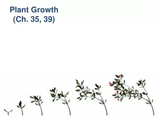
Ch 35. The Nervous System. The levels of organization in a multicellular organism include Tissues – groups of similar cells that perform a single function Organ – a group of tissues that work together to perform a complex function

E N D
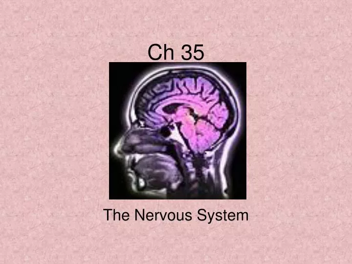
Ch 35 The Nervous System
The levels of organization in a multicellular organism include Tissues – groups of similar cells that perform a single function Organ – a group of tissues that work together to perform a complex function Organ System – group of organs that perform closely related functions Organization of the Body
Body Tissues • Four major types of tissues include • Epithelial – lines body cavities and covers body surfaces • Connective – provides support for the body and connects its parts • Muscle – contracts to allow movement • Nerve Tissue – transmits impulses to coordinate body system
Homeostasis – maintaining a controlled, stable environment Negative Feedback – a stimulus produces a response that opposes the original stimulus Positive Feedback – a stimulus produces a response that enhances the original stimulus Maintaining Homeostasis
The Nervous System • Controls and coordinates functions throughout the body and responds to internal and external stimuli. • Cells that transmit the impulses are called neurons. • Sensory Neurons – carry information to the brain and spinal cord. • Motor Neurons – carry information away from the brain and spinal cord. • Interneurons – relay messages between sensory and motor neurons.
Cell Body – main part; contains nucleus and cytoplasm; most metabolic activity occurs here Dendrites – short, branched extensions that carry impulses toward the cell body Axon – long extension that carries impulses away from the cell body Myelin Sheath – fatty layer that covers many axons; insulates the neuron and speeds up the rate of impulses. Nodes of Ranvier – gaps in the myelin sheath; impulses jump from node to node Parts of a Neuron
Nerve Impulses: Resting Potential • At rest, a neuron has a high concentration of K+ ions inside the cell. • A rest, a neuron has a high concentration of Na+ ions outside the cell. • Negatively charged proteins and Cl- ions are found inside the cell. • Resting Potential: the inside is negative with respect to the outside.
Nerve Impulses: Action Potential • A stimulus will cause the membrane to change its permeability. • The membrane becomes very permeable to Na+ and Na+ rushes in. • The membrane then becomes permeable to K+ and K+ rushes out. • The cell becomes less negative on the inside. • This is the “action potential”. • The impulse is self-propagating (causing the next point along the membrane to be activated).
Impulse strength is always the same. The minimum level of a stimulus required to activate a neuron is the threshold. Any stimulus stronger than threshold will produce an impulse. Any stimulus weaker than threshold will produce no impulse. A nerve impulse will produce an impulse or it will not produce an impulse (All-or-none principal). The Threshold of a Stimulus
Synapses • A synapse is the place where an axon meets dendrites, a muscle cell, or a gland cell. • The end of a neuron (axon tip) releases chemical messengers called neurotransmitters. • Neurotransmitters cause an impulse to travel to the next cell.
Central Nervous System(CNS) – brain and spinal cord Peripheral Nervous System (PNS) – cranial and spinal nerves. Autonomic Nervous System (ANS) – regulates involuntary activities Divisions of the Nervous System
Cerebrum – largest part Two hemispheres Site of intelligence, learning, and judgment Folds and grooves on surface increase surface area Divided into lobes Frontal Parietal Temporal Occipital The Brain
Cerebellum Coordinates and balances actions of the muscles Thalamus Receives messages from all sensory receptors and relays the information to the proper region of the cerebrum. Cerebellum and Thalamus
Hypothalamus, Medulla, Pons • Hypothalamus • Control center fro recognition and analysis of hunger thirst, fatigue, anger, body temperature • Medulla Oblongata • Contains vital reflex centers (swallowing, breathing, heart rate) • Pons • Contains important respiratory centers that affect normal breathing reflex
Link between brain and rest of body Reflex – quick, automatic response to a stimulus Allows body to respond to danger immediately; does not require brain! Reflex Arc: Receptor Sensory Neuron Interneuron Motor Neuron Affector (muscle, gland) Spinal Cord & Reflex Arc
Made up of cranial nerves and spinal nerves Sensory Division – carries info from sense organs to CNS Motor Division – carries info from CNS to muscles and glands Peripheral Nervous System
Division of PNS Two subdivisions Sympathetic Division – speeds up body process; “fight-or-flight” response Parasympathetic Division – slows down body processes Autonomic Nervous System
Ch 36 Skeletal, Muscular, and Integumentary Systems
Protect vital organs Store minerals Makes blood Aids movement (provides site for muscle attachment) Framework for body Functions of the Skeletal System
Periosteum – tough layer of surrounding connective tissue Osteocytes – bone cells Haversian Canals – provide means for blood vessels to get to bone cells Bone Marrow – soft tissue Yellow Marrow (fatty) Red Marrow (makes blood cells) Cartilage – reduces friction between bones at a joint Compact bone Spongy bone Bone Structure
Embryonic skeleton starts off as bone Eventually, it is replaced by bone tissue Process called ossification Ossification completed between ages of 18 – 25. Bone Development Human Fetus 6 Weeks
Axial Division Skull, Ribs, Sternum, Vertebrae Appendicular Division Shoulders, Arms, Hips, Legs Use Diagram to Learn Bones Bones of the Skeleton
Place where two bones meet Immovable Joints – “fixed” joints; allow no movement; held by sutures or tightly with connective tissue Slightly Movable Joints – slight movement Freely Movable Joints Ball-and-Socket Pivot Hinge Joints Joints
Miscellaneous (Bones) • Ligaments – hold bones together at a joint • Osteoporosis – weakening of bones • Arthritis – inflammation of a joint • Strain – damaged ligaments
Skeletal Muscle Has striped appearance Voluntary Found attached to bones Cardiac Muscle Has striped appearance Involuntary Found only in heart Smooth Muscle Has no striped appearance Involuntary Found in walls of hollow organs Types of Muscle Tissue
Muscle Structure • Myosin filaments • Thick proteins • Actin filaments • Thin proteins • Muscles contract because myosin pulls on actin. • Requires ATP
Neuromuscular Junction – where neuron and muscle cell meets Acetylcholine – neurotransmitter released by neurons that triggers muscle contraction Muscles can only pull on bones; not push! Muscle Contraction
Tendons – connect muscles to bone Use diagram to learn muscles Biceps brachii Triceps brachii Rectus femoris Biceps femoris Rectus abdominis Pectoralis Trapezius Gastrocnemius Gluteus Maximus Deltoideus Muscle Names
Integumentary System includes skin, hair, nails, sweat glands, etc. Functions Barrier against infection and injury Regulate body temperature Remove waste Protect against UV radiation Makes Vitamin D The Skin
Epidermis Outer layer Outermost cells are dead and keratinized Keratin is a protein that makes cells flat and water resistant Inner cells are living, contain pigment called melanin Does not contain nerve cells or blood cells. Layers of the Skin
Layers of the Skin • Dermis • Inner layer • Lies beneath epidermis • Contains blood vessels, nerves, glands, hairs • Two types of glands • Sebaceous Glands – oil glands • Sweat glands
Hair covers almost every body surface Protects body and insulates Hairs grow from the root Nails grow from nail root Fingernails grow about 4X faster than toenails. Hair and Nails
Ch 37 Circulatory and Respiratory Systems
Circulates gases like oxygen and carbon dioxide Circulates heat Circulates water Circulates nutrients, hormones, wastes Blood is always red! Functions of the Circulatory System
Plasma (55%) – yellowish Mostly water Contains dissolved nutrients, gases, wastes, etc. Corpuscles (45%) Erythrocytes – red cells; carry oxygen; hemoglobin Leukocytes – white cells; immunity Thrombocytes – platelets; coagulation Blood Composition
Arteries Smaller diameter than veins Higher BP than veins Carry blood away from heart Veins Larger diameter than arteries Lower BP than arteries Carry blood toward heart Capillaries One cell thick Only site for diffusion Blood Vessels
See p. 944 Circulation of Blood
There are two masses of nerve/muscle tissue in the heart that control the heart beat. Both are located in the right atrium. The SA Node (sinoatrial Node) – natural pacemaker; initiates beat The AV Node (Atrioventricular Node) – causes ventricles to contract Heart rate varies with activity Heart rates averages 70 beats/minute The Heart Beat
Like any pump, the heart produces pressure. When the heart contracts, it produces a wave of fluid pressure in the arteries. Blood pressure decreases when the heart relaxes. Device called sphygmomanometer measures BP. The first number is the systolic pressure (pressure when ventricles contract) The second number is the diastolic pressure (pressure when ventricles relax) Normal reading 110 – 120/80 mmHg Blood Pressure
Atherosclerosis – fatty deposits called plaque build up on the inner walls of arteries High Blood Pressure – Also called hypertension; increases risk of heart attack or stroke Heart Attack – blood flow to heart muscle blocked; heart muscle dies; symptoms include shortness of breath, nausea, chest pain Diseases of the Circulatory System
The basic function is to bring about the exchange of oxygen and carbon dioxide between the blood, air, and tissues. The respiratory system consists of the nose, pharynx, larynx, trachea, bronchi, and lungs. The Respiratory System
Parts of the Respiratory System • Nose • Hairs • Smell receptors • Mucus glands • Pharynx • Trachea • Epiglottis • Cartilage supports • Bronchi, Bronchioles • Alveoli
Gas Exchange & Breathing • Gas exchange occurs only via the alveoli. • Inhalation • Diaphragm • Ribs & Sternum • Volume vs Pressure • Exhalation • Diaphragm • Ribs & Sternum • Volume vs Pressure
Control of Breathing • Rate of breathing controlled by medulla oblongata. • Average breathing rate at rest 12 – 15 breaths per minute. • Affected by • Amount of CO2 in blood • Emotions • Exercise
Nicotine is a stimulant that increases heart rate and blood pressure. Carbon Monoxide is a poisonous gas that blocks the transport of oxygen by hemoglobin in the blood. It decreases the blood’s ability to supply oxygen to its tissues. Tar has been shown to cause cancer. Nicotine and CO paralyze the cilia of the airways. Smoking causes the lining of the airway to swell, reducing air flow. Emphysema is the loss of elasticity in the tissues of the lungs. Tobacco & The Respiratory System