Download
1 / 57
570 likes | 736 Views
MICROBIAL GENETICS. CHAPTER 7. A fair amount of the material in this chapter is repeat stuff you have had. Because of the importance of the material in chapters 7 and 8 I will cover it. Please bear with me.

E N D
MICROBIAL GENETICS CHAPTER 7 A fair amount of the material in this chapter is repeat stuff you have had. Because of the importance of the material in chapters 7 and 8 I will cover it. Please bear with me.
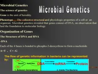
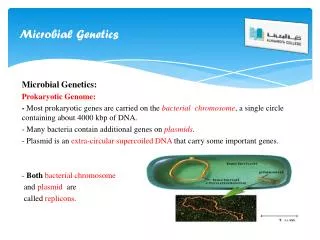
Two strands are held together by hydrogen bonding. A-T; G-C pair. If G were opposite T they would not hydrogen bond and you would get a bulge or bubble in the DNA (important in both repair and in DNA synthesis when the wrong nucleotide is accidentally added). The strands are antiparallel and the DNA molecule is twisted into a double helix. The two sugar-phosphate strands run in opposite (antiparallel) directions. Each new strand grows from the 5' end toward the 3' end (that is, nucleotides are added to the 3’ end). Fig. 7.1 The structure of DNA
You will be responsible for recognizing the different bases found in nucleic acids Figure 2.22 The five bases found in nucleic acids
Three methods of information transfer: 1. Replication- DNA makes new DNA 2. Transcription- DNA makes RNA-initial step in protein synthesis and synthesis of ribonucleoproteins (such as ribosomes) and transfer RNA. 3. Translation- RNA links amino acids together to form proteins In both DNA replication and transcription , DNA serves as a template for synthesis of a new nucleotide polymer. The sequence of bases in each polymer is complementary to that in the original DNA. In RNA, thymine is replaced by uracil, which pairs with adenine. DNA replication, transcription, and translation all transfer information from one molecule to another. These processes allow information in DNA to be transferred to each new generation of cells and to be used to control the functioning of cells. Fig. 7.3 The transfer of information from DNA to protein
http://highered.mcgraw-hill.com/sites/0072437316/student_view0/chapter14/animations.htmlhttp://highered.mcgraw-hill.com/sites/0072437316/student_view0/chapter14/animations.html How DNA nucleotides are added during DNA replication
Replication fork DNA polymerase cannot joint 2 DNA bases together it requires a primer made by another enzyme (DNA primase) http://highered.mcgraw-hill.com/sites/0072437316/student_view0/chapter14/animations.html sliding clamp and DNA polymerase fall off the DNA and reattach at the point of synthesis of the newly replicated Okazaki fragment. DNA replication fork Fig. 7.4b (this figure is not in your text but the process is described in your text). It is effectively a close-up look at Fig. 7.4.
- DNA replication in procaryote- DNA strands separate, and replication begins at a replication fork on each strand. As synthesis proceeds, each strand of DNA serves as template for the replication of its partner. The strands are antiparallel. http://www.youtube.com/watch?v=NHKh08wMrM4 Fig. 7.4 DNA replication in a prokaryote
Transcription • DNA to RNA • One strand only serves as RNA template • RNA polymerase • Promoter • Simultaneous transcription and translation
Sigma factor This figure is of prokaryotic transcription In eukaryotic cells there are a number general transcription factors instead of the sigma factor RNA polymerase binds to one and only one strand of exposed DNA-termed the “sense” strand. More specifically the RNA polymerase binds to a region of the sense strand called the promoter region. In prokaryotes, transcription and translation both take place in the cytoplasm whereas in eukaryotes, transcription takes place in the nucleus. Fig. 7.5 The transcription of RNA from template DNA. Sigma factor must be attached to the RNA polymerase to begin transcription. The RNA polymerase binds to a region “upstream of the start site of transcription
In prokaryotic cells one mRNA molecule corresponds to one or more genes and there are no introns In both the pro- and eukaryotic genes RNA polymerase binds, at the promoter region (which involves TATA binding region), to only one strand of exposed DNA-termed the "sense" strand. In eukaryotic genes one mRNA corresponds to only one gene and there are introns. However alternative splicing can result in more than 1 protein/gene. Eukaryote Fig. 7.6 Eukaryotic genes differ in complexity from prokaryotic genes
goes through a tunnel in the 50S subunit The small (30S) and large (50S) subunits are shown from two different angles. The subunits enfold the mRNA strand. The region of peptide synthesis is the junction of these three compoenents. The growing polypeptide chain passes through a tunnel in the 50S subunit, which can be seen in cross-section.Eukaryotic ribosomes are comprised of a 60S and a 40S subunit, Fig. 7.7 Prokaryotic ribosomal structure
RNA Types • Ribosomal RNA (rRNA) • Messenger RNA (mRNA) • Transfer RNA (tRNA)
Messenger RNA (mRNA). In prokaryotic cells one mRNA molecule corresponds to one or more genes (in eukaryotes one mRNA corresponds to one gene). Each mRNA molecule becomes associated with one or more ribosomes. At the ribosome, the information coded in mRNA acts during translation to dictate the sequence of amino acids in the protein. In translation each triplet (sequence of three bases) in mRNA constitutes a codon. Codons are the “words” in the language of nucleic acids. Each codon specifies a particular amino acid or acts as a terminator codon. Start codon is the first codon in the mRNA and is the codon AUG for methionine in eukaryoties and formylmethionine in prokaryotes. The last codon to be translated in a molecule of mRNA is a terminator, or stop codon. It causes the RNA polymerase to fall off the mRNA.
Nonsense codon Start codon In Mycoplasma sp. UGA codes for tryptophan- we (in my lab.) are trying to express a mycoplasmal gene (arginine deiminase) that has seven UGA’s in Escherichia coli-(the above is the genetic code that E.coli uses) what do we need to do??? Fig. 7.8 The genetic code, with standard three-letter abbreviations for amino acids
Translation • mRNA codons • tRNA anticodons • Amino acid links • Role of ribosomes
Transfer RNA (tRNA) The function of transfer RNA (tRNA) is to transfer amino acids from the cytoplasm to the ribosomes for placement in a protein molecule. Many different kinds of tRNA’s have been isolated from the cytoplasm of cells. Each tRNA has a three-base anticodon that is complementary to a particular mRNA codon
on the mRNA The two-dimensional structure of the tryptophan transfer RNA. The amino acid attaches to CCA (all tRNA’s have the same amino acid attachment site) http://www.youtube.com/watch?v=T54Akbu0ONU Amino acids are attached to the tRNA by aminoacyl-tRNA synthetase. Only the anticodon/codon is involved in the recognition of the tRNA and not the amino acid. Fig. 7.9 Transfer RNA
Another class of RNA’s have been discovered and is termed RNAi The Mechanism of RNA Interference (RNAi) Long double-stranded RNAs (dsRNAs; typically >200 nt) can be used to silence the expression of target genes in a variety of organisms and cell types (e.g., worms, fruit flies, and plants). In mammalian cells, introduction of long dsRNA (>30 nt) initiates a potent antiviral response, exemplified by nonspecific inhibition of protein synthesis and RNA degradation. The implication for use of RNAi in disease therapy are amazing and several biotech companies have been established just to exploit these possibilities. http://www.youtube.com/watch?v=e2dnFBnFWT8&feature=related Shorter version RNAi http://www.youtube.com/watch?v=UdwygnzIdVE&feature=related Longer version
Introduction of a long dsRNA RNase III-like enzyme called Dicer (initiation step). Then, the siRNAs assemble into endoribonuclease-containing complexes known as RNA-induced silencing complexes (RISCs), unwinding in the process. The siRNA strands subsequently guide the RISCs to complementary RNA molecules, where they cleave and destroy the cognate RNA (effecter step). Cleavage of cognate RNA takes place near the middle of the region bound by the siRNA strand. The Mechanism of RNA Interference (RNAi)
RNAi is another type of functional RNA The three types of RNA: rRNA, mRNA, and tRNA Fig. 7.10 Transcription and translation
Translation • mRNA codons • tRNA anticodons • Amino acid links • Role of ribosomes
Initiation of prokaryotic protein synthesis http://highered.mcgraw-hill.com/olcweb/cgi/pluginpop.cgi?it=swf::535::535::/sites/dl/free/0072437316/120077/micro06.swf::Protein Synthesis Animation of prokaryotic translation, i.e., protein synthesis
Previous slide illustrates the initiation of protein synthesis. Following the binding of the intiator codon the process proceeds as in step 3 below. The energy that drives the ribosome along the mRNA comes from GTP. E-site Translation: Protein synthesis uses 80-90% of the bacterial cell energy Fig. 7.12 Protein synthesis – Steps 1-4
the energy to make the peptide bond is in the aminoacyl tRNA E P A Fig. 7.14 Protein synthesis Steps 5-7
Feedback inhibition, also called end-product inhibition, the end product of a biosynthetic pathway directly inhibits the first enzyme in the pathway.
Regulation of Metabolism • Feedback Inhibition (Enzymatic) The synthesis of threonine involves five enzymatically controlled reactions (arrows) and four intermediate products (A,B, C, and D). Threonine (the end product) inhibits an allosteric enzyme (1) that catalyzes Reaction 1. The allosteric enzyme is functional when its allosteric site is not occupied and is nonfunctional when the end product of a sequence of reactions is bound to that site. Fig. 7.13 Feedback inhibition
Enzyme Induction. Some enzymes are maintained at comparable levels at all times in a cell. These enzymes are termed constitutive enzymes. Other enzymes are induced depending on the presence or absence of a nutrient; these are called inducible enzymes. The nutrient itself acts as an inducer of enzyme production.
Lac operon http://www.youtube.com/watch?v=iPQZXMKZEfw&feature=related
Lactose is absent the operon is “off” Lactose is present the operon is “on” Fig. 7.14 Enzyme induction (negative regulation because binding of the element blocks transcription)
cAMP-CRP (cyclic AMP-cyclic AMP receptor protein) In the presence of glucose the the lac operon is off because glucose decreases the level of cyclic AMP (cAMP) in the cell and cAMP is needed to activate the cAMP-CRP which is an necessary positive regulator of the lac operon cAMP-CRP is an example of positive regulation (binding of the element is needed for the start of transcription)
Unlike what is observed in the lac operon the addition of tryptophan causes the repressor to bind and to shut off the pathway. Unlike the lac operon in the presence of tryptophan this regulatory system shuts off. Enzyme repression
Enzyme Repression animation- Tryptophan operon http://highered.mcgraw-hill.com/olcweb/cgi/pluginpop.cgi?it=swf::535::535::/sites/dl/free/0072437316/120080/bio26.swf::The Tryptophan Repressor
Catabolite repression: A Slightly different kind of repression operates in connection with some catabolic pathways, i.e., catabolite repression. When certain bacteria E. coli,, for example, are grown of a nutrient medium containing both glucose and lactose they grow at a logarithmic rate as long as glucose is available. When the glucose is depleted, they enter a stationary phase but soon begin to grow again at a logarithmic rate, though not quite as rapidly. This time the logarithmic growth rate results from the metabolism of lactose. The stationary phase is the period during which the enzymes needed to utilize lactose are induced.
The figure above shows the growth pattern of E.coli in medium containing both glucose and Lactose Fig. 7.15 Catabolite repression. In E. coli grown in the presence of glucose the level of cyclic AMP is markedly reduced.
Point mutation-involves base substitution, or nucleotide replacement, in which one base is substituted for another at a specific location in a gene. http://www.youtube.com/watch?v=kp0esidDr-c&feature=related
http://www.youtube.com/watch?v=kp0esidDr-c Figure 7.16- The effects of base substitution (a point mutation)
Frameshift mutation is a mutation in which there is a deletion or an insertion of one or more bases. http://www.youtube.com/watch?v=e-xEQ05ncMo Frameshift mutation
http://www.youtube.com/watch?v=HYS6EKnQcv0&feature=related Repair of point mutations
There is one major question that remains: How does the mismatch repair system know the difference between strand contains the correct base and the one on which it should make the incision? The way that it tells the two strands apart is by a marker that is added to the parent strand during replication. An additional methyl (-CH3) added to the adenine base groups of the parent strand and acts as a flag for the mismatch repair system so that it knows to make its cuts on the opposite strand. Figure %: Methylated Adenines Found on the Parent Strand Only
Fig. 7.19 Acridine, a chemical mutagen. Insertion of acridine orange Into DNA helix can produce a frameshift
Thymine dimers: formation and repair http://highered.mcgrawhill.com/olcweb/cgi/pluginpop.cgi?it=swf::535::535::/sites/dl/free/0072437316/120082/micro18.swf::Thymine Dimers
The repair of DNA damage Light repair or photoreactivation , occurs in the presence of visible light in bacteria previously exposed to UV light. Dark repair, a defective segment of DNA is cut out and replaced. Some human skin cancers, such as xeroderma pigmentosum are caused by a defect in the cellular DNA repair mechanism
Light enzyme is activated by visible light Dark http://highered.mcgrawhill.com/olcweb/cgi/pluginpop.cgi?it=swf::535::535::/sites/dl/free/0072437316/120082/micro18.swf::Thymine Dimers Figure 7.21 Thymine dimer reparis repeat of the thymine dimer slide