Download
1 / 56
580 likes | 864 Views
NEMATODES -2-. Doç.Dr.Hrisi BAHAR. Hookworms of medical importance & Trichinella spiralis Filarial worms W.bancroftii. Doç.Dr.Hrisi BAHAR. Ancylostoma and Necator (Hookworms ). Causative agents of ancylostomosis and necatorosis (hookworm infection) Ancylostoma duodenale

E N D
NEMATODES -2- Doç.Dr.Hrisi BAHAR
Hookworms of medical importance&Trichinella spiralisFilarial wormsW.bancroftii Doç.Dr.Hrisi BAHAR
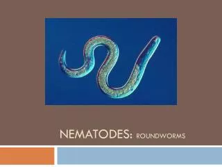
Ancylostoma and Necator (Hookworms) Causative agents of ancylostomosis and necatorosis (hookworm infection) Ancylostoma duodenale Necator americanus are common parasites of the human small intestine, causing enteritis and anemia. Infection is mainly by the percutaneous route.
Ancylostoma and Necator (Hookworms) Occurrence. ●Human hookworm infections are most frequent in the subtropics and tropics area in southern Europe, Africa, Asia,Southern US, Central and South America. ● In central Europe, hookworm infections are seen in travelers returning from the tropics or in guest workers from southern countries.
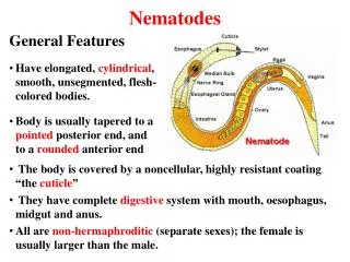
Morphology The hookworms that parasitize humans are 0.7–1.8 cm long with the anterior end bent dorsally in a hooklike shape Ancylostoma and Necator (Hookworms)
Ancylostoma The entrance to the large buccal capsule is armed with toothlike structures Ancylostoma and Necator (Hookworms) Ancylostoma duodenale
Necator The entrance to the large buccal capsule is armed with cutting plates Ancylostoma and Necator (Hookworms) Necator americanus
Ancylostoma and Necator (Hookworms) ● The thin-shelled, oval eggs (about 60µm long)containing only a small number of blastomeres are shed with feces. In one to two days the first-stage larvae leave the egg molt twice, and develop into infective third-stage larvae.
Ancylostoma and Necator (Hookworms) ● Since the shed second-stage cuticle is not entirely removed, the third-stage larva is covered by a special sheath. ● Larvae in this stage are sensitive to dryness.
Ancylostoma and Necator (Hookworms In moist soil or water they remain viable for about one month. Higher temperatures (optimum: 20–30 8C) and sufficient moisture is favor for the development of the parasite stages outside of a host.
Life Cycles of Hookworms ● Humans are infected mainly by the percutaneous route. ● Factors favoring infection include working in rice paddies, walking barefoot on contaminated soil.
Life Cycles of Hookworms 1-In favorable conditions of moisture, temperature, and oxygen, eggs develop in the soil and hatch when they reach maturity. 2-They release a rhabditiform larva which feeds for a short time and molts twice before becoming an infective filariform larva.
Life Cycles of Hookworms • Filariform larva enters the host either by being swallowed or through hair follicles.
Life Cycles of Hookworms 3-While penetrating the skin, the larvae shed their sheaths and migrate into lymphatic and blood vessels. 2-Once in the bloodstream, they migrate via the right ventricle of the heart and by tracheal migration into the small intestine, where they develop to sexual maturity.
Life Cycles of Hookworms ● Following oral infection, immediate development in the intestine is probably possible , without the otherwise necessary migration through various organs. ● The parasites can survive in the human gut for one to 15 years.
Ancylostoma and Necator (Hookworms Clinical manifestation ● Hookworms are bloodsuckers. The buccal capsule damages the mucosa and induces inflammatory reactions. ● The intestinal tissue damage results in diarrhea with bloody admixtures, steatorrhea, loss of appetite, nausea, flatulence, and abdominal pains.
Ancylostoma and Necator (Hookworms General symptoms 1-Iron deficiency anemia due to constant blood loss 2-Edemas caused by albumin losses 3-Weight loss due to reduced food uptake and malabsorption. 4-Blood eosinophilia is often present. Mild infections cause little of clinical note.
Ancylostoma and Necator (Hookworms Diagnosis. ●Diagnosis relies on finding of eggs in stool samples. ● The eggs are thin-shelled and oval ● When fresh they contain only two to eight blastomeres ● The eggs in older stool samples have already developed a larger number of blastomeres
Eggs of Hookworms Hookworm eggs
Ancylostoma and Necator (Hookworms Therapy and control Drugs effective against hookworms are pyrantel, mebendazole,and albendazole. Practicable preventive and control measures include mass chemotherapy of the population in endemic regions, reduction of dissemination of hookworm eggs by adequate disposal of fecal matter and reduction of percutaneous infection by use of properly protectiv footwear
Nematodal Infections of Tissues and the Vascular System Filarial nematodes Trichinella
Filariae The nematode genera of the superfamily Filarioidea will be here under the collective term filariae, and the diseases they cause are designated as Filarioses. In the life cycle of filariae infecting humans, insects (mosquitoes, blackflies, flies etc.) function as intermediate hosts and vectors. Filarioses are endemic in subtropical and tropical regions; in other regions they are observed as occasional imported cases.
Filariae The most important filariosis is onchocercosis, the causative agents is Onchocerca volvulus,is transmitted by blackflies. Microfilariae of this species can cause severe skin lesions and eye damage, even blindness. Diagnosis of onchocercosis is based on clinical symptoms,detection of microfilariae in the skin and eyes, as well as on serum antibody detection.
Filariae Other forms of filarioses include lymphatic filariosis .The causative agent is Wuchereria bancrofti, Brugia species and Loaosis that the causative agent is Loa loa. Dirofilaria species from animals can cause lung and skin lesions in humans
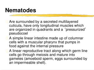
Filariae ● Filariae are threadlike nematodes. ● The length of the adult stages of the species that infect humans varies between 2–50 cm whereby the females are larger than the males. ● The females release embryonated eggs or larvae called microfilariae. ● The eggs are about 0.2–0.3 mm long, surrounded by an extended eggshell ● They can be detected in the skin or in blood
Filariae • ● Based on the periodic appearance of microfilariae in peripheral blood, periodic filaria species are differentiated from the nonperiodic ones showing continuous presence. • ● The periodic species produce maximum microfilaria densities either at night (nocturnal periodic) or during the day (diurnal periodic). • ● Different insect species, active during the day or night, function as intermediate hosts accordingly to match these changing levels of microfilaremia.
Life Cycle of Filariae Insect: Ingestion of microfilaria with a blood meal development in thoracic musculature with two moltings to become infective larva migration to mouth parts and tranmission into skin of a new host through puncture wound during thenext blood meal.
Life Cycle of Filariae Human Migration to definitive localizations and further development with two more moltings to reach sexual maturity.
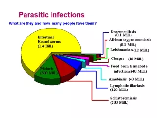
Wuchereria bancroftiCausative agents of lymphatic filariosis About 120 million people in 80 countries suffer from lymphatic filariosis caused by Wuchereria bancroftii or Brugia species (One-third each in India and Africa, the rest in southern Asia, Pacific region,and South America),
Wuchereria bancrofti Life cycle and epidemiology ●Humans are the only natural final hosts of W. bancrofti. ●The intermediate hosts of W. bancrofti are various diurnal or nocturnal mosquito genera.
Wuchereria bancrofti ● The development of infective larvae in the insects is only possible at high environmental temperatures and humidity levels; ● In Wuchereria bancrofti the process takes about 12 days at 28 ºC.
Wuchereria bancrofti ● Following a primary human infection,the filariae migrate into lymphatic vessels where they develop to sexual maturity. ●Microfilariae appear in the blood after seven to eight months.
Wuchereria bancrofti ●Pathogenesis and clinical manifestations. The initial symptoms can appear as early as 1 month , although in most cases the incubation period is 5 to 12 months or much longer. Asymptomatic infection,is with microfilaremia that can persist for years.
Wuchereria bancrofti Acute symptomatic infection: Inflammatory and allergic reactions in the lymphatic system caused by filariae Swelling of lymph nodes, lymphangitis, general malaise, swellings on egs, arms, scrotum, orchitis.
Wuchereria bancrofti Chronic symptomatic infection Chronic obstructive changes in the lymphatic system hindrance or blockage of the flow of lymph and dilatation of the lymphatic vessels (“lymphatic varices”) Indurated swellings caused by connective tissue proliferation in lymph nodes, extremities (especially the legs, “elephantiasis”), the scrotum. thickened skin ,lymphuria, when lymph vessels rupture. This clinical picture develops gradually in indigenous inhabitants over a period of 10–15 years after the acute phase, in immigrants usually faster.
Wuchereria bancrofti Tropical, pulmonary eosinophilia Syndrome with coughing, asthmatic pulmonary symptoms, high-level blood eosinophilia, lymph node swelling and high concentrations of serum antibodies (including IgE) to filarial antigens. No microfilariae are detectable in blood, but sometimes in the lymph nodes and lungs. This is an allergic reaction to filarial antigens
Wuchereria bancrofti • 1-A diagnosis can be based on clinical symptoms (frequent eosinophilia) • 2-Finding of microfilariae in blood (sampling at night for nocturnal periodic species). • 3-Microfilariae of the various species can be differentiated morphologically in stained blood smears and byDNA analysis.
Wuchereria bancrofti • 4-Detection by ultrasonography, • particularly in the male scrotal area. • 5-Detection of serum antibodies (group-specific antibodies, specific IgE and IgG subclasses) and circulating antigens • 6-The recent development of a specific ELISA and a simple quick test (the ICT filariosis card test ıs usefull for serology.
Wuchereria bancrofti • Therapy. Both albendazole and diethylcarbamazine have been shown to be at least partially effective against adult filarial stages. • However, optimal treatment regimens still need to be defined.
Wuchereria bancrofti ● In 1997, the WHO initiated a program to eradicate lymphatic filariosis.The control measure is mass treatment of populations in endemic areas with microfilaricides. ● Concurrent single doses of albendazole with either ivermectin or diethylcarbamazineare 99% effective in removing microfilariae from the blood for 1 year after treatment with measure of mosquito bites.
TrichinellaCausative agent of trichinellosis • ● Humans can acquire an infection with larvae of various Trichinella species by ingesting raw meat (from pigs, wild boars, horses, and other species). • ● Adult stages develop from the larvae and inhabit the small intestine, where the females produce larvae that migrate through the lymphatics and bloodstream into skeletal musculature, penetrate into muscle cells and encyst.