Download
1 / 51
510 likes | 641 Views
THE NERVOUS SYSTEM: NEURAL TISSUE. Nervous system functions . 1. sensory function sensory receptors detect internal and external stimuli information is sent to CNS via sensory (afferent) neurons within sensory nerves 2. integrative function

E N D
Nervous system functions • 1. sensory function • sensory receptors detect internal and external stimuli • information is sent to CNS via sensory (afferent) neurons within sensory nerves • 2. integrative function • integrates = processing of information within the CNS • stores info and also makes decisions once info is processed • one important integrative function = perception • processed by interneurons within the CNS • 90% of the neurons within the CNS are interneurons • 3. motor function • decision usually manifests itself as a motor command – contraction of a muscle, secretion by a gland • motor commands travel along motor (efferent) neurons within motor nerves • commands are sent to effectors = muscles and glands
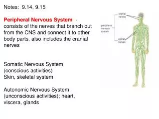
Nervous system includes all neural tissue in body • about 3% of the total body weight • Central Nervous System • Brain and spinal cord (brain = 100 billion neurons, SC = 100 million neurons) • Peripheral Nervous System • All neural tissue outside CNS • includes the spinal and cranial nerves
A schematic of the vertebrate nervous system Figure 21-6
Cells in Nervous Tissue • Neurons • Neuroglia
Neuroglia (Glia) • “glue” • about half the volume of cells in the CNS • smaller than neurons • 5 to 50 times more numerous • do NOT generate electrical impulses • divide by mitosis • however, mature glial astrocytes may not be able to divide – only precursors to glial populations • regulate the clearance of neurotransmitters • participate in neural development • provide growth factors and chemical cues for the development of neurons and their axonal processes • Two types in the PNS • Schwann cells • satellite cells • Four types in the CNS • Astrocytes • Oligodendrocytes • Microglia • Ependymal cells
Astrocytes • Largest of glial cells • Most numerous • Star shaped with many processes projecting from the cell body -two types: protoplasmic, fibrous -protoplasmic – short branches, found in gray matter -fibrous – many long unbranched processes, found in white matter -processes make contact with the capillaries supplying the CNS, the neurons of the CNS and the pia mater membrane covering the brain and spinal cord • Help form and maintain blood-brain barrier • processes wrap around the blood capillaries and isolate the neuron from the blood supply -also secrete substances that maintain a unique permeability for the endothelial cells that line these capillaries – restricts movement of substances out of the blood • Provide structural support for neurons • microfilaments within cytoskeleton • Maintain the appropriate chemical environment for generation of nerve impulses/action potentials • Regulate nutrient concentrations for neuron survival • Regulate ion concentrations - generation of action potentials by neurons • Take up excess neurotransmitters – take up excess GABA and glutamate • Assist in neuronal migration during brain development • Perform repairs to stabilize tissue – scar formation???
Oligodendrocytes • Each forms myelin sheath around the axons of neurons in CNS • Analogous to Schwann cells of PNS • Form a supportive network around CNS neurons • fewer processes than astrocytes • round or oval cell body
Microglia • few processes • derived from mesodermal cells • that also give rise to monocytes • and macrophages • Small cells found near blood vessels • 15% of the glial cells of the CNS • Phagocytic role - clear away dead cells • derived from hematopoietic stem cells • protect CNS from disease through phagocytosis of microbes • migrate to areas of injury where they clear away debris of • injured cells - may also kill healthy cells
Ependymal Cells • epithelial cells arranged in a • single layer • range in shape from cuboidal • to columnar • Form epithelial membrane lining cerebral cavities (ventricles) & central canal - that contain CSF • Produce & circulate the cerebrospinal fluid (CSF) found in these chambers • CSF = colourless liquid that protects the brain and SC against • chemical & physical injuries, carries oxygen, glucose and other necessary • chemicals from the blood to neurons and neuroglia
PNS: Satellite Cells • Flat cells surrounding PNS axons • Support neurons in the PNS • help regulate the chemical environment surrounding the neurons
PNS: Schwann Cells • each cell surrounds multiple unmyelinated PNS axons with a single layer of its plasma membrane • Each cell produces part of the myelin sheath surrounding an axon in the PNS • contributes regeneration of PNS axons
Neurons • what is the main defining characteristic of neurons? • have the property of electrical excitability - ability to produce • action potentials or impulses in response to stimuli
Representative Neuron http://www.horton.ednet.ns.ca/staff/selig/Activities/nervous/na1.htm • -neurofilaments or neurofibrils give cell shape and support - bundles of • intermediate filaments • -microtubules move material inside cell • -lipofuscin pigment clumps (harmless aging) - yellowish brown • -the processes that emerge from the body of the neuron = nerve fibers • -two kinds: dendrites & axons • 1. cell body or soma (or perikaryon) • -single nucleus with prominent nucleolus (high synthetic activity) • -Nissl bodies • -rough ER & free ribosomes for protein synthesis • -proteins then replace neuronal cellular components for growth and repair of damaged axons in the PNS
Neurons 2. Cell processes = dendrites (little trees) - the receiving or input portion of the neuron -short, tapering and highly branched -surfaces specialized for contact with other neurons -cytoplasm contains Nissl bodies & mitochondria
3. Cell processes = axons • Conduct impulses away from cell body-propagates nerve impulses to another neuron • Long, thin cylindrical process of cell • contains mitochondria, microtubules & neurofibrils - NO ER/NO protein synth. • joins the soma at a cone-shaped elevation = axon hillock • first part of the axon = initial segment • most impulses arise at the junction of the axon hillock and initial segment = trigger zone • cytoplasm = axoplasm • plasma membrane = axolemma • Side branches = collaterals arise from the axon • axon and collaterals end in fine processes called axon terminals • Swollen tips called synaptic end bulbs contain vesicles filled with neurotransmitters
Axonal Transport • Cell body is location for most protein synthesis • neurotransmitters & repair proteins • however the axon or axon terminals require proteins • e.g. neurotransmitters • Axonal transport system moves substances • slow axonal flow • movement of axoplasm in one direction only -- away from cell body • movement at 1-5 mm per day • replenishes axoplasm in regenerating or maturing neurons • fast axonal flow • moves organelles & materials along surface of microtubules • at 200-400 mm per day • transports material in either direction • for use in the terminals or for recycling in cell body
Axonal Transport & Disease • fast axonal transport route by which toxins or pathogens reach neuron cell bodies • tetanus (Clostridium tetani bacteria) – toxin = tetanospasmin • disrupts motor neurons causing painful muscle spasms • “lockjaw” – muscle stiffness usually involves jaw and neck first • interferes with the release of neurotransmitters that result in inhibition of muscle contraction • neuronal targets are peripheral motor end plates, CNS, sympathetic NS • lethal dose = 2.5 ng per kg body weight (e.g. 70 ng for 175 lbs) • bacteria enter the body through a laceration or puncture injury • more serious if wound is in head or neck because of shorter transit time to the brain
Structural Classification of Neurons • Based on number of processes found on cell body • multipolar = several dendrites & one axon • most common cell type in the brain and SC • bipolar neurons = one main dendrite & one axon • found in retina, inner ear & olfactory • unipolar neurons = one process only, sensory only (touch, stretch) • develops from a bipolar neuron in the embryo - axon and dendrite fuse and then branch into 2 branches near the soma - both have the structure of axons (propagate APs) - the axon that projects toward the periphery = dendrites
Structural Classification of Neurons • Named for histologist that first described them or their appearance • Purkinje = cerebellum • Renshaw = spinal cord • others are named for shapes • e.g. pyramidal cells
Functional Classification of Neurons • Sensory (afferent) neurons • transport sensory information from skin, muscles, joints, sense organs & viscera to CNS • Motor (efferent) neurons • send motor nerve impulses to muscles & glands • Interneurons (association/integrative) neurons • connect sensory to motor neurons • 90% of neurons in the body
Terms to know • membrane potential = electrical voltage difference measured across the membrane of a cell • resting membrane potential = membrane potential of a neuron measured when it is unstimulated • results from the build-up of negative ions in the cytosol along the inside of the neuron’s PM • the outside of the PM becomes more positive • this difference in charge can be measured as potential energy – measured in millivolts • polarization • depolarization • repolarization • hyperpolarization
ion channels in the PM of neurons and muscles contributes to their excitability • when open - ions move down their concentration gradients • channels possess gates to open and close them • two types: gated and non-gated Ion Channels • 1. Leakage (non-gated) or Resting channels: are always open, contribute to the resting potential • -nerve cells have more K+ than Na+ leakage channels • -as a result, membrane permeability to K+ is higher • -K+ leaks out of cell - inside becomes more negative • -K+ is then pumped back in • 2. Gated channels: open and close in response to a stimulus • A. voltage-gated: open in response to change in voltage - participate in the AP • B. ligand-gated: open & close in response to particular chemical stimuli (hormone, neurotransmitter, ion) • C. mechanically-gated: open with mechanical stimulation
The resting potential, generated mainly by open “resting”, non-gated K+ channels -the number of K+ channels dramatically outnumbers that of Na+ -however, there are a few Na leak channels along the axonal membrane ECF AXON
Graded potentials • local changes in membrane potential that occur in varying intensities (grades) • caused by the opening of ion channels in a region of the axonal membrane • usually ligand-gated or mechanically-gated channels • typically gated ion channels for sodium – results in a slight depolarization = graded potential • region that is being depolarized = active area • stronger the triggering event = stronger the graded potential that results • the stronger the trigger the more ion channels open, the greater the depolarization • spread by passive current flow • because a local area has begun to depolarize – charge of this area changes • specifically the inside area gets more positive in relation to the surrounding areas that are at rest • the outer area becomes more negative in relation to the surrounding areas that are at rest • this produces a current that starts to spread to the surrounding areas – depolarizing them • BUT they die over short distances • this current decreases as it travels further from the originating area
Action Potential • Resting membrane potential is -70mV • triggered when the membrane potential reaches a threshold usually -55 MV • if the graded potential change exceeds that of threshold – Action Potential • Depolarization is the change from -70mV to +30 mV • Repolarization is the reversal from +30 mV back to -70 mV) • action potential = nerve impulse • takes place in two stages: depolarizing phase (more positive) and repolarizing phase (more negative - back toward resting potential) • followed by a hyperpolarizing phase or refractory period in which no new AP can be generated http://www.blackwellpublishing.com/matthews/channel.html
depolarization (increase in MP) results from opening of Na+ channels. This opens an increasing number of voltage-gated Na channels which depolarizes the membrane more. Once threshold is reached, a large # of voltage-gated Na+ channels open and a rapid increase in MP results outflow of K+ restores the resting MP. Na+ channels begin to open and K+ channels close. K+ outflow results in hyperpolarization (below resting) results in a refractory period. at a certain stage of depolarization, theMP also opens voltage-gated K+ channels which permit the outflow of K+ . The Na+ close and the MP becomes more negative returning toward resting MP
Local Anesthetics • Prevent opening of voltage-gated Na+ channels • Nerve impulses cannot pass the anesthetized region • Novocaine and lidocaine – blocks nerve impulses along nerves that detect pain
Continuous versus Saltatory Conduction • Continuous conduction (unmyelinated fibers) • An action potential spreads (propagates) over the surface of the axolemma http://highered.mcgraw-hill.com/sites/0072437316/student_view0/chapter45/animations.html#
Saltatory Conduction • Saltatory conduction • -depolarization only at nodes of Ranvier - areas along the axon that are unmyelinated and where there is a high density of voltage-gated ion channels • -current carried by ions flows through extracellular fluid from node to node http://www.blackwellpublishing.com/matthews/actionp.html
Rate of Impulse Conduction • Properties of axon • Presence or absence of myelin sheath • Diameter of axon • The propagation speed of a nerve impulse is not related to stimulus strength. • Larger = faster conduction • Myelin 5-7 X faster • larger, myelinated fibers conduct impulses faster due to size & saltatory conduction
Myelination increases the velocity of impulse conduction Figure 21-15
Action Potentials in Nerve and Muscle • Entire muscle cell membrane versus only the axon of the neuron is involved • Resting membrane potential • nerve is -70mV • skeletal & cardiac muscle is closer to -90mV • Duration • nerve impulse is 1/2 to 2 msec • muscle action potential lasts 1-5 msec for skeletal & 10-300msec for cardiac & smooth • Fastest nerve conduction velocity is 18 times faster than velocity over skeletal muscle fiber
Synapse • Synapse • Site of intercellular communication between 2 neurons or between a neuron and an effector (e.g. muscle) • Permits communication between neurons and other cells • Initiating neuron = presynaptic neuron • Receiving neuron = postsynaptic neuron • Most are axodendritic axon -> dendrite • Some are axoaxonic – axon > axon • axon terminal swell to form synaptic end bulbs or form swollen bumps called varicosities • release of neurotransmitters from synaptic vesicles • multiple types of NTs can be found in one neuron type http://www.lifesci.ucsb.edu/~mcdougal/neurobehavior/modules_homework/lect3.dcr
Synapses • NTs will cause either and excitatory or inhibitory response • If the NT depolarizes the postsynaptic neuron = excitatory • Often called an excitatory postsynaptic potential (EPSP) • Opening of sodium channels or other cation channels (inward) • Some NTs will cause hyperpolarization = inhibitory • Often called an inhibitory postsynaptic potential (IPSP) • Opening of chloride channels (inward) or potassium channels (outward) • Neural activity depends on summation of all synaptic activity • Excitatory and inhibitory
Synapses • Chemical • Membranes of pre and postsynaptic neurons do not touch • Synaptic cleft exists between the 2 neurons – 20 to 50 nm • the electrical impulse cannot travel across the cleft – indirect method is required – chemical messengers (neurotransmitters) • Most common type of synapse • The neurotransmitter induces a postsynaptic potential in the PS neuron – type of AP • Communication in one direction only http://www.blackwellpublishing.com/matthews/nmj.html
Chemical synapse • Is the conversion of an electrical signal (presynaptic) into a chemical signal back into an electrical signal (postsynaptic) • 1. nerve impulse arrives at presynaptic end bulbs • 2. fusion of synaptic vesicles to PM - role for calcium • 3. release of NTs • 4. opening of channels in PM of postsynaptic neuron (e.g. sodium) • 5. postsynaptic potential develops – depolarization & triggering of AP in postsynaptic neuron
Chemical synapse • propagation of AP at the target post-synaptic neuron usually involves opening of ligand-gated Na+ channels on the membrane of the post-synaptic neuron • binding of NT to a receptor on post-synaptic membrane • this receptor is the ligand-gated channel
Release of NTs from Synaptic end bulbs Synaptic vesicles can be filled, exocytosed, and recycled within a minute • -synaptic vesicles are filled with NTs • the vesicles move into proximity near the PM of the end bulb = active zone • -upon receipt of AP into these bulbs -causes the opening of voltage-gated Ca2+ channels • -the influx of calcium promotes the • “docking” of the synaptic vesicle with the PM and the exocytosis of their contents • -the synaptic vesicle components • are recycled for future use
Synapses • Electrical • Direct physical contact between cells required • Conducted through gap junctions • Two advantages over chemical synapses • 1. faster communication – almost instantaneous • 2. synchronization between neurons or muscle fibers • e.g. retina, heart-beat
Neurotransmitters • More than 100 identified • Some bind receptors and cause channels to open • Others bind receptors and result in a second messenger system • Results in either excitation or inhibition of the target • Removal of NTs • 1. Diffusion • move down concentration gradient • 2. Enzymatic degradation • e.g. acetylcholinesterase • 3/ Uptake by neurons or glia cells • neurotransmitter transporters • e.g. NE, epinephrine, dopamine, serotonin
1. small molecules: Acetylcholine (ACh) • All neuromuscular junctions use ACh • ACh also released at chemical synapses in the PNS and by some CNS neurons • Can be excitatory at some synapses and inhibitory at others • Inactivated by an enzyme acetylcholinesterase • Blockage of the ACh receptors by antibodies = myasthenia gravis - autoimmune disease that destroys these receptors and progressively destroys the NMJ • Anticholinesterase drugs (inhibitors of acetylcholinesterase) prevent the breakdown of ACh and raise the level that can activate the still present receptors
Neurotransmitters 2. Amino acids: glutamate & aspartate & GABA • Powerful excitatory effects • Glutamate is the main excitatory neurotransmitter in the CNS • Stimulate most excitatory neurons in the CNS (about ½ the neurons in the brain) • Binding of glutamate to receptors opens calcium channels = EPSP • GABA (gamma amino-butyric acid) is an inhibitory neurotransmitter for 1/3 of all brain synapses
GABA • GABA action is affected by a broad range of drugs called benzodiazepines • e.g. lorazepan – Ativan • e.g. diazepam - Valium • Various uses: hynoptic, sedative, anxiolytic, anticonvulsant, muscle relaxant, amnesic • Short lasting – half life is less than 12 hours • hypnotic effects • insomnia • Long lasting – half life is more than 24 hours • anxiolytic effects (anti-anxiety drug) • Acts to enhance GABA • GABA – major inhibitory NT in the CNS • GABA binds to GABA receptors – several types • Benzodiazepines bind and modulate the activity of the GABAA receptor which is the most prolific NT receptor in the brain • GABAA receptor is comprised of 5 protein subunits • One subunit is the alpha subunit • BZ’s bind to the alpha subunit only and increase its affinity for binding the GABA neurotransmitter • The GABAA receptor is a ligand-gated chloride channel • Binding of GABA increases the inward flow of chloride ions which hyperpolarizes the neuron and inhibits its ability to make a new action potential • Therefore BZ’s potentiate the inhibitory effects of GABA
Valium • top selling drug from 1969-1982 • GABA agonist • Also decreases the synthesis of neurosteroid hormones (e.g. DHEA, progesterone) which may regulate emotional state • Acts on areas of the limbic system, the thalamus and the hypothalamus (anti-anxiety drug) • Metabolized by the liver into many metabolites • Gives rise to a biphasic half live of 1-2 days and 2-5 days! • Lipid-soluble and crosses the blood-brain barrier very easily • Stored in the heart, the muscle and the fat • Some drugs (barbituates), anti-depressants and alchohol can enhance its effect • Smoking can increase the elimination of valium and decrease its effects
Neurotransmitters 3. Biogenic amines: modified amino acids • catecholamines:norepinephrine (NE), epinephrine, dopamine (tyrosine) • serotonin - concentrated in neurons found in the brain region = raphe nucleus • derived from tryptophan • sensory perception, temperature regulation, mood control, appetite, sleep induction • feeling of well being • NE - role in arousal, awakening, deep sleep, regulating mood • epinephrine (adrenaline) - flight or fight response • dopamine - emotional responses and pleasure, decreases skeletal muscle tone Other types: a. ATP - released with NE from some neurons b. Nitric oxide - formed on demand in the neuron then release (brief lifespan) -role in memory and learning -produces vasodilation
Dopamine • Involved in feelings of pleasure, strength • Also mediates skeletal muscle contraction • Neurotransmitters like dopamine, serotonin, glutamate, acetylcholine etc… are secreted and then rapidly internalized by transporters in order to control their levels within the nervous system • Many drugs affect these transporters • Ritalin = methylphenidate • Stimulant used to treat ADD, ADHD, narcolepsy amd chronic fatigue • 1954 – initially prescribed for depression and narcolepsy • 1960 – prescribed to children with ADD, ADHD • Reason?? Might be due to an imbalance in dopamine • Binds both dopamine and norepinephine transporters and inhibits their ability to take these NTs back up (keeps their levels high in the synapse) • Dopamine transporters (DAT) found in the PM of neurons (presynaptic) • Transports dopamine back into the neuron along with sodium ions (symporter) • This terminates the dopamine signal • Chloride ions are also required to enter the neuron to prevent depolarization • In adults – these transporters regulate dopamine levels • Cocaine – binds and inhibits DATs – increasing dopamine in the synapse • Amphetamines – binds amphetamine receptors on a neuron which causes the internalization of the DAT into the neuron – increasing dopamine in the synapse
Neuropeptides • widespread in both CNS and PNS • excitatory and inhibitory • act as hormones elsewhere in the body • -Substance P -- enhances our perception of pain • -opioid peptides: endorphins - released during stress, exercise • -breaks down bradykinins (pain chemicals), boosts • the immune system and slows the growth of cancer • cells • -binds to mu-opioid receptors • -released by the neurons of the Hypothalamus and by • the cells of the pituitary • enkephalins - analgesics • -breaks down bradykinins (200x stronger than morphine) • -pain-relieving effect by blocking the release of • substance P • dynorphins - regulates pain and emotions • **acupuncture may produce loss of pain sensation because of release of opioid-like substances such as endorphins or dynorphins