Download
1 / 44
440 likes | 452 Views
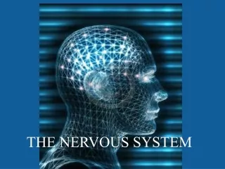
THE NERVOUS SYSTEM (SYSTEMA NERVOSUM). THE FUNCTIONS OF THE NERVOUS SYSTEM. controling function The neural cont r ol of the activity of the organs The basical property is the excitability and the generating of electric nerve impulses. The nerve cell (neuron).

E N D
THE FUNCTIONS OF THE NERVOUS SYSTEM controling function • The neural control of the activity of the organs • The basical property is the excitability and the generating of electric nerve impulses
The nerve cell (neuron) • the basic component of the nervous system • highly specialized (excitability, conductivity) Glial cell (neuroglia) • support, nutritive, defensive and other functions
Neuron • the body (perikaryon) • the projections • dendrites: receiving of complaints • Axon (nerve fibre): conducts electrical impulses away from the neuron's cell body
The types of the neurons • Multipolar: the largest ones, several dendrites and one axon, star-shaped, 80% • Pseudounipolar dendrite + axon - dendraxon, T-shaped, central and peripheral branch (spinal ganglions and ganglions of cranial nerves) • Unipolar only one projection - axon (primary sensory cells, olfactory cells, rods and cones of the retina) • Bipolar one axon and one dendrite, (the second neuron of the optic tract)
The synapse (a part of axon) • Ended by enlargement– terminal button – is in contact with other neuron – the connection synapse (muscle – neuromuscular junction) • presynaptic membrane, postsynaptic membrane • Transfer of impulse– spread through axon centrifugally as electric signal – action potential • terminal button – vesicles with neurotransmitters
Neuroglia • Astrocytes: the largest ones long unbranched cellular processes (with vascular feet), which cover all vessels of CNS – blood-brain barrier • Oligodendrocytes: within the grey and white matter, they myelinate nerve fibres of CNS (nutrition, homeostasis) • Microglia: the smallest ones (primarilly within the bone marrow) defense function – ability of phagocytosis) • Ependymal cells:they cover the central canal of spinal cord and the brain ventricles –they are bathed by the cerebrospinal fluid and they help its flow
Neuroglia Function: they accelerate the conduction of the excitement, nutrition, homeostasis, defence function
The myelin sheath CNS: oligodendroglia PNS: Schwann cells • insulator, it is interrupted by the nodes of Ranvier, the neural impulses ,,jump´´ • Myelinized nerve fibres - quicker transmission of the impulse The bodies of the neurons: ganglions, grey matter of CNS The projections of the neurons: white matter of CNS, nerves of PNS
THE DIVISION OF THE NERVOUS SYSTEM 1. central nervous system(systema nervosum centrale) Spinal cord (medulla spinalis) brain (encephalon, cerebrum) • The hind-brain (rhombencephalon) • The Medulla oblongata(medulla oblongata) • The pons (pons Varoli) • The cerebellum (cerebellum) • The midbrain (mesencephalon) • The fore-brain (prosencephalon) • The diencephalon (diencephalon) • The cerebrum (telencephalon) 2. peripheral nervous system(systema nervosum periphericum) It connects CNS with the periphery of organism (centripetally, centrifugally) • Spinal nerves (nervi spinales) • Cranial nerves (nervi craniales) • sympathicus(pars sympathica) • parasymphaticus(pars parasympathica) Cerebrospinal nerves Autonomic nerves
The terms: • funiculus = cord x • fasciculus= bundle of axons -HETEROGENEOUS structure, it originates within the different nuclei of the grey matter and creates synapses also within the different structures x • tractus= tract – bundle of axons - HOMOGENOUS structure, the fibres have a common origin and end • rostral
The nervous system 1)It mediates relations between external environment and organism 2) It provides body´s response to stimuli from outside 3) It mediates relations between all parts of organism 4) It provides integrality of all actions within organism
FUNCTIONAL TYPES OF AXONS WITHIN PNS somatosensory Skin perception, proprioception, pain viscerosensory mechanoception, pain Afferent taste, hearing and vestibular informations, senzory somatomotor striated muscle striated muscle branchiomotor Efferent Smooth muscle visceromotor sympathetic myocardium parasympathetic glands
THE REFLEX ARC The reflex is a fyziologic action, whose essence isthe response of the organismto the change of the external or the internal environment- this is a response of the organismto theirritation. The nervous system constantly monitorsthe conditions of the external and internal environment of the organism using the receptors, it processes these informationswithin CNS andgives off theinstructions for the executive organs – effectors = REFLEX The anatomical underlay of the reflex is thereflex arc – a system of the neural tracts, on which is the reflex carried out. 1. Receptors 2. Afferentneural tracts 3. CNS 4. Efferrent neural tracts 5. Effectors
RECEPTORS Receptor (sensor):responds to the changes of the internal or external environment of the organism and it converts them into action potential of the neural impulses, these impulses are emited intodirecting centre within CNS The division according to the location: exteroreceptors – receptors, which respondtothe stimuli (changes) from the external environment of the organism interoreceptors– receptors, which respondtothe stimuli (changes) from the internal environment of the organism According to the specific location: proprioreceptors – receptors which are located within the locomotor system (within themuscles, tendons andarticular capsules) visceroreceptors – receptors which are located within the visceral organs and the walls of the vessels
The division according tothe physical character of the acting stimulus mechanoreceptors – receptorsreponding to mechanicalstimuli chemoreceptors – receptorsreponding tochemicalstimuli thermoreceptors – receptorsreponding tothermalstimuli photoreceptors – receptorsreponding tolightstimuli The special case present so calledalgoreceptors – receptors which respond to pain. The muscles • Muscle and tendon spindles (intrafusal fibres) – degree of contraction and tension of the muscle fibers • Free nerve ending around the muscle fibers - pain
The afferent nerve pathways The afferent (centripetal, sensory)pathways conduct the neural impulsesfrom the receptorsintoCNS. They are formed by the projections of sensory neurons, whose bodies are located out of CNS withinsensory ganglions. The afferent pathways are divided into: Somatosensory pathways – bring information from receptors within skin and locomotor system Viscerosensory pathways – bring informations from visceroreceptors (from visceral organs) Sensory pathways – bring informations from sensors – specialized sensory organs (visual, auditory, vestibular, olfactory and gustatory sytems)
THE CENTRAL NERVOUS SYSTEM The central nervous system (CNS) is the directing centre of the nervous system. It takes the informationsfrom the receptorsusingthe afferent nerve pathways.It processes and evaluates these informations andprovidesthe responsesof the organism usingthe efferent nerve pathways and effectors. The nervous tissue of CNS isformed by two types of the matter: The grey matter (substantia grisea) The white matter(substantia alba)
The grey matter (substantia grisea): is formed by the bodies and dendrites of the neurons + glial cells The bodies of the neurons take the informationsfrom axons of sensory neuronsof the sensory ganglions andgive offthe new informationsfor motor neurons within CNS On the connection between sensory and motor neuronsmay not be any otherswitching neuron (by the simpliest reflexes), but usually there are one ore more switching neurons, so called interneurons, inserted
The white matter (substantia alba): Is formed by the bundles of myelinized axons of neurons, which are located within the grey matter and provide the communication between the neuronsof the grey matter Associationpathways Commissuralpathways Projectionpathways – according to the direction, we divide them into two groups: Ascending tracts – tracts passing from the lower centre toward the higher one.They are the continuation of the afferent peripheral tracts. Descendingtracts – tracts passing from the higher centre toward the lower one. They are the continuation of the efferent peripheral tracts
The efferent nerve pathways The efferent, centrifugal, motor) nervepathways: They lead the nervous impulses from CNS into effectors (executive organs) They begin withthe motor neuronwithin CNS, axon of this neuron leaves CNS and presents the proper efferent (motor) pathway Somatomotor pathways Visceromotor pathways
EFFECTORS Effectors are the executive organs or tissues, which provide the proper response of the organism to the irritation. They can be: Muscle cells– the result of the reflexu is the motion Glandular cells– the result of the reflexu is the secretion
THE PERIPHERAL NERVOUS SYSTEM(systema nervosum periphericum) The periphery nervous system is formed by a system of nerves and ganglions, whichconveyreversibletransfer of the informationsbetweenCNS and the periphery (skin, locomotor system andvisceral organs) Nerveis consist of nervefibres andfibrous connective tissue Nervefibresare myelinated (white) orunmyelinated (grey) projections of the neurons Nervefibrescreatefascicles, a few fascicle create nerve, Particular nervefibresare interconnected using the fibrous connective tissue – endoneurium, within the nerve the fascicles of the fibres are interconnecte usingperineurium Teh surface of the nerve is covered by fibrous layer– epineurium
Sensory nerves • They consist the afferent fibres, which lead the information from the receptors toward CNS • Within theirs course, there are sensory ganglions inserted with the bodies of pseudounipolar neurons • Peripheral branch leads the impulses from receptors within the periphery into the ganglion, the bundles of these peripheral branches form the nerve • Central branch leads the impulses from the ganglion toward the grey matter of CNS somatosensory– lead the informations from the receptors within the skin and the locomotor system (muscles, tendons, periosteum, articular capsules) viscerosensory –lead the information from receptors within the organs sensory – lead the informations from sensors
motor nerves • They consist the efferent fibres, which leadfrom CNS into effectors (muscles or glands) andprovide theiractivity • They originate within the nuclei of the grey matter of the spial cord or the brainstem (body of the neuron) somatomotor nerves – innervatestriated muscle, the nerve fibers (axons) enter the muscles directly without switch-over visceromotor nerves – innervatesmooth muscleandglandularcells, into which the nerve fibers enter after at least one switch-over within autonomic ganglion Mixed nerves • They consist both motor and sensory fibers. The most of the nerves are mixed, only some are either clearly motor or clearly sensory
The peripheral system 1. According to from which part of CNS the nerves arise, we can divide them into: The cranial nerves (pass through the base skull) The spinal nerves(rise through foramina intervertebralia) 2. According to the innervated parts of the body: somatic nervous system • It is controlled by our consciousness • It is consist of somatosensory (or sensory) and somatomotor nerve fibers (tracts) • It does the sensory innervation of the skin and the locomotor system (muscles, tendons, bones, articular capsules) and the motor innervation of the striated muscle autonomic nervous system • It works independently on our consciousness • sympathicus, parasympathicus
THE SPINAL NERVES (nervi spinales) • The spinal nerves rise from the spinal cord in the number of 31 pairs The cervical nerves(nervi cervicales) – 8 pairs The thoracic nerves(nervi thoracici) – 12 pairs The lumbar nerves(nervi lumbales) – 5pairs The sacral nerves(nervi sacrales) – 5pairs The coccygeal nerve(nervus coccygeus) – 1pair • Each spinal nerverises from the spinal cord through two roots, the anterior one (radix ventralis) and the posterior one (radix dorsalis) • The anterior roots consist only efferent fibres (motor), the posterior roots only afferent fibres(sensory) • Within the course of the posterior root, there is the spinal ganglion(ganglion spinale)inserted
The posterior root (radix dorsalis) consists afferent fibres, which serve to superficial and deep perception, leading of pain, warmth and cold. On each posterior root, there is ganglion spinale located, which consists neurons of the afferent fibers. The anterior root (radix ventralis) consists efferent somatomotor fibres (for striated muscle) and visceromotor fibres (for smooth muscle cells within the walls of organs and vessels, the skin etc.).
The proper spinal nerv is formed by connection of the anterior and posterior root and it rises from the spinal canal throughforamen intervertebrale After the leaving of the spinal canal, the spinal nerve divides into 2 branches – the posterior branch (ramus dorsalis)andthe anterior branch (ramus ventralis), both branches consist the afferent and efferent tracts.
Ramus dorsalis: doesn´t form plexuses Ramus ventralis: forms plexuses The nerve plexuses are allways formed only by the anterior branches of the appropriate spinal nerves!
The posterior branches (rami dorsales) –don´t form the plexuses They are short andthinbranches of the spinal nerve function: motor and sensory innervation of the back part of the trunk (mixed nerves) They do the motor innervation of autochtoneí (deep) back muscles and sensory innervation of the skin in adjacent area During their course, they keep simple segmentalarrangement Only the posterior branches of C1 – C3, L1 – L3 a S1 – S3have more complicated arrangement
C1 – C3 n. suboccipitalis n. occipitalis major n. occipitalis tertius • suboccipital muscles + skin L1 – L3 a S1 – S3 nn. clunium superiores nn. clunium medii • skin of the gluteal region
Ramus dorsalis: • ramus medialis (deep back muscles and the skin of medial part of the back) • ramus lateralis (deep back muscles in the lateral part of the back)
The anterior branches (rami ventrales) • They are longer and thicker branches of the spinal nerve • Function: motor and sensory innervation of the anterior part of the trunk • They do the motor innervation ofthe muscles of the anterior part of the trunk (neck except infrahyoid muscles, thorax, abdomen and palevis) and the muscles of the limbs and the sensory innervation of the skin of the anterior part of the trunk and the limbs • They have more complicated arrangement than the posterior branches of the spinal nerves, they lost the segmentation and created nerve plexuses): • Plexus cervicalis C1-C4 • Plexus brachialis C4-Th1 • Nervi thoracici Th1-Th12 (they keep segmental arrangement) • Plexus lumbalis Th12-L4 • Plexus sacralis L4-5,S1-5,Co
THE CERVICAL PLEXUS(plexus cervicalis) • It is created by the anterior branches of first four cervical nerves (C1 – C4) • It is located sides the cervical vertabral column and several sensory, motorandmixed nerve rise from it. The particular nerves of the cervical plexus innervate: Sensory innervation:the skin in the neck area, a part of the head and a part of upper extremity girdle Motor innervation:neck muscles (infrahyoid muscles, musculi scaleni anddeep neck musclesanddiaphragm, which originates in the neck area during embryonic development) • suprahyoid muscles + m. sternocleidomastoideus + m. platysma are of gill origin an they are innervated by some cranial nerves, not cervical nerves
The cervical plexus The sensory branches – they rise together by the posterior margin of m. sternocleidomastoideus – punctum nervosum sensitivum: n. occipitalis minor (C2–3) n. auricularis magnus (C2-C3) ramus anterior et posterior n. transversus colli (C3)- rr. superiores et inferiores- ansa cervicalis superficialis nn. supraclaviculares (C3-4) mediales intermedii laterales
The motor branches – for pre- and intervertebral muscles, m. scalenus medius, m. sternocleidomastoideus, m. trapezius a m. levator scapulae
Ansa cervicalis (profunda) (C1–3) – (ansa n. hypoglossi) motor fibres forinfrahyoid muscles, exceptm. thyrohyoideus (separate branch of n. thyrohyoideus) • radix sup. (C1) – joins n. hypoglossus and passes between a. carotis comm. and v. jugularis int. • radix inf. (C2–3) - along v. jugularis int., above tendon of m. omohyoideus both roots connect and ansa (loop) is created
The mixedbranches n. phrenicus (C3–5) Itdivides on theinferiorsurfaceofthediaphragm – rr. phrenicoabdominales – motorinnervationofthediaphragm, sensoryinnervationofthe peritoneum, gallbladder, pancreas; r. pericardiacus – sensory innervationofthepericardium and adjacent part ofpleura nn. phrenici acc. – accessoryfibres a) directlyfromplx. brachialis (C5–6), b) through n. subclavius c) friomradix inf. ansaecervicalis Theycan substitute typicaln. phrenicus Irritationofthenerve – singultus (hiccup)
THE THORACIC NERVES(nervi intercostales) • They keep segmentalarrangement, don´t form plexuses, pass within intercostal spaces from behinf forwardtogetherwith the intercostal vessels (within sulcus costae on the inferior margin of the rib) • 1. – 6. toward the sternum, 7. – 12. toward the anterior abdominal wall they innervates: • Sensory i.:skin of thorax and abdomen andparietalsheetof pleura and peritoneum • Motor i.: properthoracic (intercostal) muscles and anteriro and lateralgroup of abdominal muscles
Nn. thoracici Th1-12 • Don´t form plexuses nn. intercostales n. subcostalis • rr. musculares (autochtone muscles of thorax) • rr. cutanei- laterales- nn. intercostobrachiales - anteriores (by the sternal margin) Skin of thorax and abdomen • rr. pleurales from Th1- strong connection into plexus brachialis (nervus intercostobrachialis)
Nn. intercostobrachiales (n. cutaneus brachii medialis)
Obrázky: • Atlas der Anatomie des Menschen/Sobotta. Putz,R., und Pabst,R. 20. Auflage. München:Urban & Schwarzenberg, 1993 • Netter: Interactive Atlas of Human Anatomy. • Naňka, Elišková: Přehled anatomie. Galén, Praha 2009. • Čihák: Anatomie I, II, III. • Drake et al: Gray´s Anatomy for Students. 2010