Download
1 / 1
10 likes | 95 Views
Brain Activation in Specific Regions in Depressed Adolescents. OBJECTIVE To use Blood Oxygen Level Dependent fMRI in conjunction with behavioral data to find differences in brain activation between

E N D
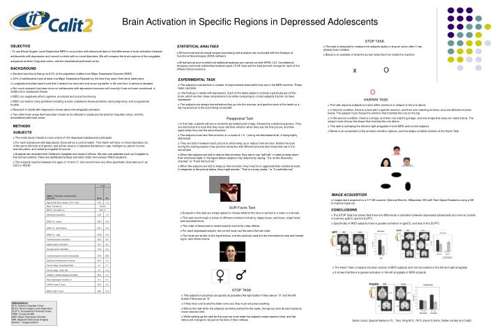
Brain Activation in Specific Regions in Depressed Adolescents • OBJECTIVE • To use Blood Oxygen Level Dependent fMRI in conjunction with behavioral data to find differences in brain activation between • adolescents with depression and normal controls with no mood disorders. We will compare the brain regions of the amygdala, • subgenual anterior Cingulate cortex, and the dorsolateral prefrontal cortex. • STOP TASK • This task is designed to measure the subjects ability to stop an action after it has already been initiated • Below is an example of what the screen looks like from inside the machine • BACKGROUND • Studies have found that up to 8.3% of the population suffers from Major Depressive Disorder (MDD) • 20% of adolescents have at least one Major Depressive Episode by the time they reach their early adult years • Longitudinal studies have found that it tends to be recurrent and occurring earlier in life now than in previous decades • Not much research has been done on adolescents with depression because until recently it has not been considered a childhood or adolescent illness • MDD can negatively effect cognitive, emotional and social functioning • MDD can lead to many problems including suicide, substance abuse problems, early pregnancy, and occupational trouble • Research in adults with depression shows abnormal amygdala activation • Two other brain areas that have been shown to be affected in adults are the anterior cingulate cortex, and the dorsolateral prefrontal cortex • STATISTICAL ANALYSES • All functional and structural images processing and analyses are conducted with the Analysis of Functional Neuroimages (AFNI) software • All behavioral and correlational statistical analyses are carried out with SPSS 14.0. Correlational Analyses examined relationship between peak LF/HF data and the total percent change for each of the different facial emotions • EXPERIMENTAL TASK • The subjects must perform a number of experimental tasks while they are in the fMRI machine. These tasks capitalize • on the findings in adults with depression. Each of the tasks seeks to activate a particular part of the brain, which we then draw comparisons from while comparing to control subjects that do not have depression • The subjects are always trained before they go into the scanner, and practice each of the tasks on a lap top previous to the scan being conducted • Reappraisal Task • In this task, subjects will see a randomly generated pixel image, followed by a disturbing picture. They are told before the scan that they must rate their emotion when they see the first picture, and then again when they see the second picture • The subjects must rate their emotion on a scale of 1-4, 1 being not distressed at all, 4 being highly distressed • They are told in between each picture to either keep up or reduce their emotion. Before the scan during the training session they practice doing this with different pictures than those that are in the actual task • When the subjects are told to reduce their emotion, they are to use “self talk” in order to keep down their emotional state. In the figure below subjects may respond by saying, “It is on the Discovery channel,” or “It will not hurt me” • When the subjects are told to keep up their emotion, they must try to aggravate their emotional state. In response to the picture below, they might answer, “That is a scary snake,” or “It could bite me” • HARRIRI TASK • This task requires subjects to match either emotions or shapes to the one above. • In the first condition, there is a face with a specific emotion, and then one matching emotion, and one different emotion below. The subject must choose the emotion that matches the one on the top • In the second condition, there is a shape, and then one matching shape, and one shape that does not match below. The subject must choose the shape that matches the one above • This task is activating the left and right amygdala in both MDD and control subjects • Below is an screenshot of the emotion condition (above), and the shape condition (below) of the Harriri Task • METHODS • SUBJECTS • The entire study intends to have a total of 100 depressed adolescents participate • For each adolescent with depression, there will be a control match. This match will have no mood disorders, be of the same ethnicity and gender, and will be close to 2 standard deviations in age, intelligence, parent income and education, and verbal and spatial IQ scores • Subjects are recruited from Children’s hospitals and doctor’s offices. We also use websites such as Craigslist to find normal controls. Fliers are distributed to Boys and Girls Clubs’ and various YMCA locations • The subjects must be between the ages of 12 and 17, and cannot have any other psychiatric disorders such as OCD or ADHD • IMAGE ACQUISITION • Images were acquired on a 3-T GE scanner (General Electric, Milwaukee, WI) with Twin Speed Gradients using a GE 8-channel head coil • GUR Faces Task • Subjects in this task are simply asked to choose whether the face or symbol is a male or a female. • The task runs through a series of different emotions including: happy faces, sad faces, angry faces and surprised faces • The order of these sets is randomized to control for order effects • For each depressed subject, the control must use the same Gur set order • The faces are similar to the figure below, and the symbols used are the international male and female signs, also shown below • CONCLUSIONS • The STOP Task has shown that there are differences in activation between depressed adolescents and normal controls in both the sgACC and the DLPFC • Specifically in MDD subjects there is greater activation in sgACC, and less in the DLPFC • The Harriri Task compares the brain activity of MDD subjects and normal controls in the left and right amygdala • It shows that there is greater activation in the left amygdala of MDD subjects • STOP TASK • The subjects must press (as quickly as possible) the right button if they see an “O” and the left button if they see an “X” • If they hear a tone and the letter turns red, they must not press anything • Before the task while the subjects are being trained for the tasks, the lap top records each subjects mean reaction time • While setting up the task for the scan we must enter the subject’s mean reaction time, and the letters will change to red just at the time of their reflexes Abbreviations: ACC: Anterior Cingulate Cortex BOLD: Blood Oxygen Level Dependent DLPFC: Dorsolateral Prefrontal Cortex FMRI: Functional MRI MDD: Major Depressive Disorder MRI: Magnetic Resonance Imaging SGACC: Subgenual ACC Sarah Jurick, Special thanks to P.I. Tony Yang M.D., Ph.D.,Kevin S Hahn, Stefan Jordan and Calit2