Download
1 / 21
400 likes | 1.15k Views
Lynsey Zuar, D.O. 30 August 2013. DDH. This describes a spectrum of conditions related to hip development in infants/children Abnormal development of acetabulum and proximal femur. The newborn hip. Newborns can often have physiologic laxity of the hip

E N D
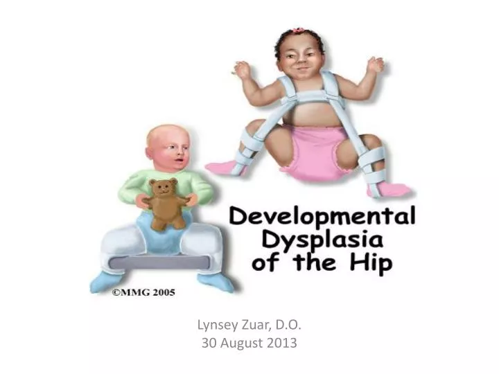
Lynsey Zuar, D.O. 30 August 2013
DDH • This describes a spectrum of conditions related to hip development in infants/children • Abnormal development of acetabulum and proximal femur
The newborn hip • Newborns can often have physiologic laxity of the hip • Can be caused by immaturity of acetabulum during first few weeks of life • DDH usually occurs in otherwise healthy infants
DDH Risk Factors • Most important 3: • Breech positioning at ≥34 weeks gestation (whether or not external cephalic version is successful) • (+) family history • Female sex • Other factors to consider: • Seen more often with first born infants • Increased incidence with oligohydramnios and related to other conditions a/w tight intrauterine packaging (eg, torticollis & metatarsus adductus)
Epidemiology • Approx 2 to 4 in 1000 live births • Prevalence rates of 1.5 in 1000 in whites • Somewhat lower in blacks • Normal left occiput anterior position in utero (which is the most common presentation during labor) places the left hip against the mom's spine and limits its abduction – 3x more common in the left hip than right.
Clinical Signs • Depends on age • Ranges from instability on newborn exam to subtle abduction of the hips • Can see an asymmetric gait in toddlers • Activity related pain in adolescents/older children • Can lead to osteoarthritis in adults • Many infants with undiagnosed/untreated hip dysplasia with begin to walk and reach other developmental milestones at the appropriate time
Klisic test: place the index finger on the ASIS and middle finger on greater trochanter. Normally the line between these points passes through or above the umbilicus if no DDH ( - test). If below the umbilicus then + Klisic test, and most likely + DDH
Ortolani & Barlow Tests • Barlow maneuver: Do this first. ADDuct hip while applying downward pressure on knee (may hear “pop”) • Ortolani maneuver: Confirms (+) above finding. Flex knee and hips to 90degr and with index finger place ant pressure on greater trochanters – aBDucting with thumbs • (+) finding is a “clunk” as femoral head relocates *Best to do this one leg at a time.
Additional Findings • Asymmetry of the gluteal thigh or labral skin folds • Decreased abduction on the affected side • Standing or walking with external rotation (‘duck-footed”) or waddling gait with hyperlordosis if b/l • Toe walking on the affected side • Leg-length inequality • Galeazzi sign: unequal knee heights
Screening • AAP recommends regular screening at all well visits until 12mo/o • If (+) Ortolani/Barlow sign is found during newborn screen the infant should be rechecked at 2wks, if still (+) then referred to an orthopedist. • If unequivocal at 2wks consider U/S at 3-4 weeks • Orthopedic referral is also recommended when the Ortolani sign is unequivocally positive (a ?clunk) • No referral needed if only softly positive finding in the examination noted(eg, hip click without dislocation) • If results of the newborn PE definitively (+) ordering an U/S is not needed as rx decisions are not influenced by the results, but are based on PE • Baby girls born breech should be screened with U/S at 6wks or radiograph of pelvis/hips at 4wks
Radiological Tests • Real-time U/S – established as an accurate method for imaging the hip in infants bc cartilage can be visualized and can also assess the morphologic features of the acetabulum. • One concern with routine U/S of newborns is increased false(+) results • Radiographs have limited value initially • Acetabular dysplasia is best found by a radiographic examination at 6mo of age or older
Treatment • The earlier it is found, the easier it is to treat, and the better outcome long term • Early – usually treated with a harness, with low risk for complications. • DDH later in childhood usually requires surgical intervention – higher risk for complications • Not diagnosed until adulthood – may have debilitating, end-stage, degenerative hip joint disease.
Nonsurgical Treatment • Newborn: Placed in a Pavlik harness for 1-2 mo – a soft positioning device that keeps the acetabulum in the socket, and help tighten the ligaments around the hip joint to promote normal hip socket formation. • 1-6mo: Baby's acetabulum repositioned into socket using a harness or similar device. • Usually successful • If not successful may have to anesthetize and move the it into proper position, and then put into a spicacast. • Triple diaper is not recommended as there is no evidence of it being beneficial
Surgical Treatment • 6mo - 2yrs: Child is placed under anesthesia, and acetabulum placed in proper position • Open surgery is sometimes necessary • Afterwards, the child is placed into spica body cast • >2yrs: Deformities may worsen, making open surgery necessary to realign, and afterwards placed into spica body cast • X-rays and other regular follow-up monitoring are needed after DDH treatment until the child's growth is complete.
Other Conditions a/w Hip Dysplasia • Teratologic hip dysplasia: • Ehlers Danlos • Down syndrome • Arthogryposis • Neuromuscular hip dysplasia: • Spina bifida • Cerebral palsy
References • American Academy of Pediatrics • American Academy of Orthopedic Surgeons • American College of Radiology • Uptodate • Medscape