Download
1 / 55
550 likes | 662 Views
Chapter 4 Skin and Body Membranes. Biology 112 Tri-County Technical College Pendleton, SC. Membranes and more…. Body membranes cover surfaces, line body cavities, & form protecting (often lubricating) sheets around organs fall into TWO major groups

E N D
Chapter 4 Skin and Body Membranes Biology 112 Tri-County Technical College Pendleton, SC
Membranes and more… • Body membranes cover surfaces, line body cavities, & form protecting (often lubricating) sheets around organs fall into TWO major groups • EPITHELIAL membranes include the CUTANEOUS, MUCOUS, and SEROUS membranes • All contain epithelial sheet always combined with underlying connective tissue • These membranes are actually simple organs
Membranes, cont. • CONNECTIVE tissue membranes represented by SYNOVIAL membranes • Two major categories of body membranes, EPITHELIAL and CONNECTIVE are classified in part according to their tissue makeup
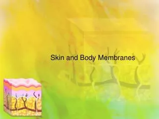
Cutaneous Membranes • Cutaneous membrane is the SKIN • Its superficial EPIDERMIS composed of keratinizing stratified squamous epithelium • Underlying DERMIS is mostly dense (fibrous) connective tissue • Unlike other epithelial membranes, CUTANEOUS membrane is exposed to air and is a DRY MEMBRANE
Mucous Membranes • Mucous membrane (mucosa) composed of epithelium resting on loose connective tissue membrane called LAMINA PROPRIA • Lines ALL body cavities open to the exterior • Hollow organs of respiratory, digestive, urinary, and reproductive tracts • Stratified squamous (mouth and esophagus) or Simple columnar (rest of digestive tract)
Mucous Membranes, cont. • “WET” or moist membranes continually bathed in secretions • Urinary mucosa bathed with urine • Mucosa epithelium adapted for ABSORPTION/SECRETION • Mucosa of respiratory/digestive tracts secrete large amounts of protective, lubricating mucus • Urinary tract DOES NOT secrete mucus
Serous Membranes • Composed of layer of simple squamous resting on thin layer of areolar connective tissue • Line ALL body cavities closed to exterior (except for dorsal body cavity and joint cavities) • SEROUS membranes occur in PAIRS • PARIETAL LAYER lines specific portion of wall of ventral body cavity then folds back on itself to form VISCERAL LAYER which covers outside of organs in cavity
Serous membranes, cont. • In body, serous membranes separated by thin, clear fluid called SEROUS FLUID that is secreted by both membranes • Allows organs to slide easily across cavity walls and one another without friction • Extremely important when mobile organs such as pumping heart and churning stomach are involved
What’s in a name? • Specific names of serous membranes depend on their locations • PERITONEUM is serous lining abdominal cavities and covering its organs • PLEURA is serosa lining the lungs • PERICARDIUM is serosa lining around the heart
Connective Membranes • Composed of connective tissue and contain NO epithelial cells • Line fibrous capsules surround joints • Provide smooth surface and secrete lubricating fluid called SYNOVIAL FLUID • Also line small sacs of CT called BURSAE and tubelike TENDON SHEATHS • Both of these cushion organs moving against each other during muscle activity (movement of tendon across a bone’s surface)
Skin and more… • Skin is major organ of integumentary system • Insulates, cushions, & protects from mechanical damage (bumps/cuts), chemical damage (acids/bases), thermal damage (heat/cold), UV radiation (sunlight), and bacteria • Uppermost (outermost) layer full of KERATIN and CORNIFIED (hardened) to prevent water loss from body surface
Skin, cont. • Capillary network and sweat glands play role in regulating heat loss from body surface • Acts as mini-excretory system: urea, salts, and water lost via sweat • Manufactures several proteins important in immunity and synthesizes vitamin D • CUTANEOUS sensory receptors (NS) located in skin • Tiny sensors include touch, pressure, temperature, and pain receptors
Skin Structure • Skin composed of two kinds of tissue • Outer EPIDERMIS=stratified squamous capable of keratinizing (becoming hard/tough) • Underlying DERMIS=dense connective tissue • Epidermis and dermis firmly connected • Burn causes separation = blister • Epidermis composed of 5 layers called STRATA • From inside out: stratum BASALE, SPINOSUM, GRANULOSUM, LUCIDUM, and CORNEUM
Skin Structure, cont. • DERMIS composed of 2 zones or layers • From inside out: RETICULAR LAYER and DERMAL PAPILLAE • Deep to dermis is the SUBCANTANEOUS TISSUE (HYPODERMIS) which is essentially adipose tissue • NOT considered part of the skin • Serves as shock absorber and insulates deeper tissues • Responsible for curves that are more a part of a woman’s anatomy than a man’s
De layer…de layer • STRATUM BASALE is deepest layer of epidermis • Lies closest to the DERMIS • Contains the ONLY epidermal cells that receive adequate nourishment (diffusion) of nutrients from dermis • Cells in this layer constantly undergoing mitosis • Also called stratum germinativum • Millions of new cells produced DAILY
Stratum Spinosum • Stratum spinosum layer situated above stratum basale and means “spiny layer” • Consists of 8 to 10 layers of cells, mostly KERATINOCYTES (most abundant epithelial cells) bound together by desmosomes • Begin to become flatter and increasingly full of keratin • Cells in this layer continue to divide
Stratum Granulosum • “Grainy” layer superficial to stratum spinosum • 3-5 layers of keratinocytes that have stopped dividing • Become more flatter and produce copious amounts of keratin • Cell membrane thickens, nuclei and organelles disintegrate • Cells die and dehydrate creating tightly interlocked layer of keratin fibers surrounded by KERATOHYALIN
Stratum Lucidum • This layer occurs ONLY where skin is hairless and extra thick • Palms of hands and soles of feet • Glassy stratum lucidum (clear layer) covers stratum granulosum • Cells in this layer are flattened, densely packed, and filled with keratin • Doomed because NOT able to get adequate nutrients and oxygen
Stratum Corneum • Found at surface of thick and thin skin • Normally 15-20 layers of keratinized cells • Keratinization (cornification) occurs in exposed skin surfaces except anterior surfaces of the eye • OMG, Darling, you are very beautiful…but • Dead cells within each layer remain tightly interconnected by desmosomes • Shed as large sheets rather than single cells • Layer is water-resistant but not waterproof • Journey from stratum basale to stratum corneum takes about 15-30 days • Dead cells remain in stratum corneum for about 2 weeks before being shed or washed away
Melanin and the Body Beautiful • Melanin is a pigment that ranges from yellow to brown to black • Produced by special cells called MELANOCYTES • Melanocytes found primarily in stratum basale • Skin exposed to sunlight, melanocytes stimulated to produce more melanin = tanning • Pigment umbrella • Freckles/moles are seen where melanin is [ ]ed in one spot
Dermis and beyond • Dermis is strong, stretchy envelope that helps hold body together • Dense fibrous connective tissue making up dermis consists of two major regions • PAPILLARY LAYER is upper dermal region • Uneven with fingerlike projections from its superior surface called DERMAL PAPILLAE which indent epidermis above • Many dermal papillae contain capillary loops which supply nutrients to epidermis
Dermis, cont. • Other dermal papillae house pain receptors (free nerve endings) and touch receptors (Meissner’s corpuscles) • On palms of hand and soles of feet, papillae arranged in definite pattern that forms looped and whorled ridges on epidermal surface that increase friction and enhance gripping ability of fingers and feet • Ridges of fingertips covered with sweat pores and leave identifying film of sweat called FINGERPRINTS on nearly everything they touch
Dermis, cont. • RETICULAR LAYER of dermis is deepest skin layer • Contains blood vessels, sweat and oil glands, and deep pressure receptors (Pacinian corpuscles) • Many phagocytes found in reticular layer • Both collagen and elastic fibers found with layer • Collagen = toughness of dermis but also attracts and binds water • Elastic = skin elasticity during youth • Aging decreases # of collagen and elastic fibers, skin begins to sag and wrinkle…tell me about it!!!!
Sidebar on Skin Colors • Three pigments contribute to skin color • Melanin, carotene, and hemoglobin • Amount and kind of melanin (yellow, reddish brown, or black) in the epidermis (basale) • Amount of carotene deposited in stratum corneum and subcutaneous tissue (yellow-orange pigment) • Amount of oxygen bound to hemoglobin (pigment in RBCs) in dermal blood vessels Example: lots of melanin = brown skin tone
Skin Colors, cont. • Light skinned people, crimson color of oxygen-rich hemoglobin in dermal blood supply shows through = “rosy glow” • Large amounts of carotene-rich food consumed, skin takes on yellow-orange cast • CYANOSIS-occurs when hemoglobin is poorly oxygenated and blood/skin of Caucasians appear BLUE • ERYTHEMA (redness)-name given reddened skin and may indicate embarrassment, fever, hypertension, inflammation, or allergy
Skin Colors, cont. • PALLOR (blanching)-certain types of emotional stress (fear, anger, & others) cause some people to become PALE • Pale skin may also signify anemia, low blood pressure, or impaired blood flow to area • JAUNDICE (yellow cast)-name given abnormal yellow skin tone; usually signifies liver disorder • Excess bile pigments absorbed into blood and deposited in body tissues • Bruises/Black and blue marks-reveal sites where blood has escaped from circulation and clotted in tissue spaces • Such clotted masses called HEMATOMAS • Unusual tendency to bruise may be indicate deficiency of vitamin C or hemophilia
Skin Appendages • Skin appendages are cutaneous glands, hair and hair follicles, and nails • Each arises from epidermis and plays unique role in maintaining homeostasis • CUTANEOUS GLANDS • Are all EXOCRINE glands that release their secretions to skin surface via DUCTS • Are two groups of cutaneous glands: • Sebaceous glands and sweat glands
Sebaceous glands • These “oil glands” are found all over body except on palms of hands and soles of feet • Ducts usually open into hair follicles but some open onto skin surface • SEBUM is lubricant that keeps skin soft and moist and prevents hair from becoming brittle • Also contains chemicals that kill bacteria • Sebaceous glands become very active when male sex hormones produced in > amounts (in both sexes) during adolescence
Sebaceous glands, cont. • Skin becomes oilier during this period of life • Sebaceous gland’s duct becomes clogged = whitehead • Accumulated material oxidizes and dries = blackhead • ACNE is active infection of sebaceous glands accompanied by “pimples” on skin • SEBORRHEA (cradle cap) in infants caused by overactivity of sebaceous glands • Scalp turns pink, raised lesions gradually form yellow to brown crust that sloughs off as oily dandrull
Sweat of one’s brow… • Two types of sweat glands: eccrine and apocrine • ECCRINE sweat glands most numerous and are found all over the body • Produce SWEAT-clear secretion that is primarily water plus some salts (sodium chloride), vitamin C, traces of metabolic wastes (ammonia, urea, & uric acid), and lactic acid (attracts mosquitoes) • Sweat is usually acidic (pH 4-6) helping inhibit growth of bacteria
Eccrine Sweat glands, cont. • Typically sweat reaches skin via duct opening externally as funnel-shaped pore • Eccrine glands play important part of body’s heat regulating equipment • Supplied with nerve endings that cause them to “sweat” when external temp or body temp is high • As sweat evaporates, carries large amounts of body heat with it • Can lose 7 liters of body water a day in very hot temps • Heat regulating functions important to maintaining life
Apocrine Glands • Apocrine glands largely confined to axillary and genital areas of body • Usually > than eccrine glands and their ducts empty into hair follicles • Secretions contain fatty acids and proteins as well as “stuff” in eccrine gland secretion • Secretion is “odorless” but bacterial growth may impart musky, unpleasant odor • Begin to function at puberty (androgens) • Play minimal role in thermoregulation • Activated by nerve fibers during pain, stress, and/or sexual foreplay/activity
Neither “hair” nor there • HAIR is produced by a root follicle and is flexible epithelial structure • HAIR ROOT is part of hair enclosed in follicle • HAIR SHAFT is part projecting from surface of skin or scalp • Hair formed by division of well-nourished cells in stratum basale epithelial cells in growth zone (hair bulb matrix) • Bulk of hair shaft, like bulk of epidermis, is dead material
Goose bumps…or whatever • Small bands of smooth muscle cells called ARRECTOR PILI connect each side of hair follicle to dermal tissue • When these muscles contract (cold, frightened, …not going there), hair is pulled upright • Dimples skin surface with “goose bumps”
If I had a hammer… • NAIL is scalelike modification of epidermis similar to hoof/claw of other animals • EACH nail has FREE EDGE, BODY (visible, attached portion), and ROOT (embedded in the skin) • Borders of nail over overlapped by skin folds called nail folds • Thick, proximal nail fold is called the CUTICLE • STRATUM BASALE of epidermis extends beneath nail as the NAIL BED
Nails, cont. • Nail’s thickened proximal area that is responsible for nail growth is called the NAIL MATRIX • LUNULA is white crescent over the thickened nail matrix • Nails, like hair and the stratum corneum cells, are mostly nonliving material
Infectious Disorders of the Skin • TINEA PEDIS (athlete’s foot)-itchy, red peeling condition of skin between toes from fungus infection • BOILS-inflammation of hair follicles and sebaceous glands, common on dorsal neck • CARBUNCLES-composite boils typically caused by bacterial infections (Staphylococcus aureus) • COLD SORES-small fluid-filled blisters that itch/sting caused by herpes simplex virus • Localizes in cutaneous nerve where dormant until activated by emotional upset, fever, or UV radiation • Usually occur around lips/oral mucosa of mouth
Skin disorders, cont. • CONTACT DERMATITIS-itching, redness and swelling of skin, progressing to blisters • Caused by exposure of skin to chemicals (poison ivy, etc.) that promote allergic responses in sensitive individuals • IMPETIGO-pink, water-filled lesions (around mouth/nose) that develop yellow crust and rupture • Caused by highly contagious staphylococcus infection • Common in elementary school children
Skin Disorders, cont. • PSORIASIS-chronic condition characterized by reddened epidermal lesions covered with dry, silvery scales • If severe, may be disfiguring • Cause unknown, but heredity seems to be implicated • Attacks often triggered by trauma, infection, hormonal changes, and stress
Three Types of Burns • BURN is tissue damage and cell death caused by intense heat, electricity, UV radiation, or certain chemicals • Burns are classified according to their severity • FIRST-DEGREE-only epidermis is damaged • Area becomes red and swollen • Temporary discomfort but “usually” NOT serious • Heal in 2-3 days without any special attention • Sunburn is “usually” a first-degree burn
Burns, cont. • SECOND DEGREE-involve injury to epidermis and upper region of dermis • Skin red/painful and BLISTERS appear • Sufficient numbers of epithelial cells still present, regrowth (regeneration) of epithelium can occur • Normally, no permanent scarring results IF infection prevented • First- and Second-degree burns referred to as PARTIAL-THICKNESS BURNS
Burns, cont. • THIRD-DEGREE-destroy entire thickness of skin and are called full-thickness burns • Burned areas appear blanched (gray-white) or blackened • Nerve endings in area destroyed so burn area NOT painful • NO regeneration is possible so skin grafting required to cover underlying exposed tissues • Considered CRITICAL if over 25% of body has second-degree burns or if over 10% of body has third-degree burns OR if there are third-degree burns of face, hands, or feet
Rule of Nines • Burns result in two life-threatening problems • Body loses supply of fluids containing proteins and electrolytes as they seep from burned surface • This dehydration/loss of electrolytes can lead to shut down of kidneys and CIRCULATORY SHOCK (inadequate circulation of blood due to low blood volume) • Volume of fluid loss can be estimated indirectly by determining how much of surface has been burned using the RULE OF NINE
Rule of Nine, cont. • Method divides body into 11 areas each accounting for 9 percent of total body surface • Additonal area surrounding genitals (perineum) = 1 percent