Download

1 / 22
220 likes | 369 Views
Getting organized – how bacterial cells move proteins and DNA. Martin Thanbichler and Lucy Shapiro Nature Reviews, 2008. Anna Buch 25.01.2010. Model systems for bacterial cell biology. E. coli : history, genetic tools, physiology B. subtilis : cell differentiation, large size

E N D
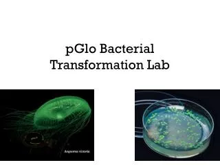
Getting organized –how bacterial cells move proteins and DNA Martin Thanbichler and Lucy Shapiro Nature Reviews, 2008 Anna Buch 25.01.2010
Model systems for bacterial cell biology • E. coli: history, genetic tools, physiology • B. subtilis: cell differentiation, large size • C. crescentus: cell division, synchonizable mobile sessile Box 1
Diffusion and capture SpoIVB SpoIIQ Assembly of stationary protein complexes Mother cell Septal membrane Phagocytosis-like uptake Figure 1
SpoIVB SpoIIIAH SpoIIQ Assembly of stationary protein complexes • Targeted membrane insertion Figure 1
Assembly of stationary protein complexes • Targeted membrane insertion Shigella flexneri: facultative intracellular pathogen IcsA: outer membrane protein, N-term is exposed to host cytoplasm IcsP: Protease that cleaves off IcsA Steinhauer et al., Mol Microbiol. 1999 32:367-77.; Pollard & Cooper, Science 2009 326:1208-12
Dynamic protein scaffolds and cell shape:Bacterial actin-like cytoskeleton Bundles of two or more protofilaments. Figure 2
MreB dynamics in C. crescentus MreB cables Spiral like during growth Ring-like during cell division Figure 2
Architecture of MreB cables B. subtilis, FRAP of GFP-Mbl Figure 2; Carballido-Lopez & Errington, Dev Cell. 2003 4:19-28.
Regulation of cell-wall biosynthesis B. subtilis Peptidoglycan (PG) synthetic machinery PG-hydrolase subunit CW binding subdomain MreB homologues: MreBH and Mbl LytE: peptidoglycan hydrolase Carballido-Lopez et al., Dev Cell. 2006 11:399-409
MreC DAPI Role of MreC in bacterial morphogenesis C. Crescentus PBC (penicillin-binding protein): involved in peptidoglykan synthesis Divakaruni et al., PNAS 2005 102:18602-7
Crescentin C. crescentus: creS::Tn5 -> no crescentin creS::Tn5 + creS ->crescentin on plasmid In-vitro assay His-CreS filaments, EM negative stain Ausmees et al., Cell. 2003 115:705-13.
Actin superfamiliy member (type II partitioning system) Walker ATPase (type I partitioning system) Tubulin homologue Plasmid segregation
Plasmid segregation by actin-like proteins Plasmid R1 of E. coli Figure 3
Walker A cytoskeletal ATPase (WACA) Plasmid segregation by Walker-type ATPases Plasmids F and pB171 of E. coli Adapted from Lim et al., PNAS 2005 102:17658-63
TubZ: B. thuringiensis serovar israelensis (pBtoxis) Plasmid segregation by a tubulin homologue E.coli, expressing TubZ-GFP, FRAP, time in sec Model proposes treadmilling Larsen et al., Genes Dev. 2007 21:1340-52
Arrangement of chromosomal DNA Figure 4, Viollier et al., PNAS 2004 101:9257-62
Divisome: Bacterial cell-division apparatus Rod-shaped bacterium (e.g. E. coli) Z-ring: FitsZ filaments Allard & Cytrynbaum PNAS 2009 106:145-50; Erickson, PNAS 2009 106:9238-43
Division-site placement: The Min system minCDE operon: MinD: WACA family MinCD-complex: inhibit FtsZ-ring formation MinE: represses MinCD activity “Fail-safe mechanism”: nucleoid occlusion B. subtilis: Noc E. coli: SlmA Figure 5
Division-site placement: The MipZ system MipZ: ATPase, inhibits FtsZ-polymerization ParB: chromosome partitioning protein parS: cluster of sites, 15 kb away from ori
Tubulin filaments: cell-division apparatus, plasmid segregation Actin cables: DNA partitioning, cell-shape determination, protein localization WACA ATPases: DNA segregation, cell-division plane Conclusions
Positioning of proteins at cell poles TipN Peptidoglycans Cardiolipin -> ProP Biochemical assembly mechanisms Actin homologues Tubulin homologues WACA ATPases Outlook