Download
1 / 43
520 likes | 1.01k Views
The Lower Extremity The Hip. ESAT 3600 Fundamentals of Athletic Training. Lower Extremity Introduction. 4 subunits Hip Knee Ankle Subtalar joint Similar in structure to the upper extremity. Lower Extremity Introduction. Difference in basic function Upper extremity Reaching Throwing

E N D
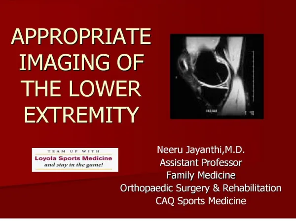
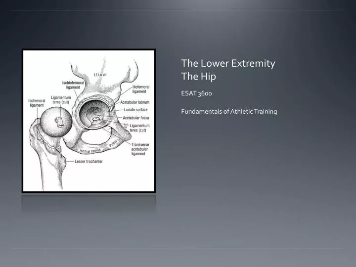
The Lower ExtremityThe Hip ESAT 3600 Fundamentals of Athletic Training
Lower Extremity Introduction • 4 subunits • Hip • Knee • Ankle • Subtalar joint • Similar in structure to the upper extremity
Lower Extremity Introduction • Difference in basic function • Upper extremity • Reaching • Throwing • Grasping • Lower extremity • Locomotion • Weight bearing
Nerves • Lumbar plexus • L1-L4 • Femoral nerve • Obturator nerve
Sacral plexus • L5-S3 • L4/S5 • Sciatic nerve • Common peroneal nerve • Tibial nerve
Pelvic Girdle • Pelvis is a segmental bridge between the vertebral column and the lower extremity • Articulations of Pelvic Girdle • Sacroiliac joint • Iliac and sacrum • Pubic symphysis
Movements of the Pelvis • Anterior tilt • Posterior tilt • Lateral tilt • Rotation
Hip Joint • Articulation of the Pelvis and femur • Acetabulum and head of femur • Ball and socket
Bony Landmarks of the Hip • Acetabulum • Pubis • Anterior superior iliac spine (ASIS) • Anterior inferior iliac spine • Illiac crest • Ischialtuberosity
Bony Landmarks of the Hip • Head of femur • Neck of femur • Great and lesser trochanter • Linea aspera • Adductor tubercle
Hip Joint Stability • VERY STABLE JOINT • Moderately strong bony arrangement • Strong ligaments • Strong muscle stability
Ligamentous Stability • Iliofemoral ligament • Y ligament • Superior band • Inferior band • Pubofemoral
Ligamentous Stability • Ischiofemoral • Posterior capsule • Triangular shape • Limits medial rotation and adduction in flexed position
Ligamentous Stability • Acetabular ligament • Transverse ligament • Ligamentum capitas femoris • AKA - Ligamentum teres • Intracapsular ligament • Links head of femur to transverse ligament
Lower Extremity Alignments–Hip • Changes in angle between femoral neck and femoral shaft throughout life span • Coxa valga • Large angle • Coxa vara • Small angle
Lower Extremity Alignments–Hip • Normal projection of femoral neck relative to femoral condyle • Adult = 8-15 degrees • Anteversion • Increased hip IR • Squinting patella • Retroversion • Increased hip ER • Feet rotated outward
Closed-packed Position of Hip • Full extension • Slight internal rotation • Slight abduction
Hip Arthrokinematics • Abduction/adduction • Roll direction of motion • Slide in opposite • Internal/external rotation • Roll direction of motion • Slide in opposite • Flexion/extension • Spin
Psoas Major • O: anterior surfaces of transverse processes, lateral borders of vertebral bodies and corresponding intervertebral discs of T12-L5 • I: lesser trochanter of femur and for short distance below along medial border of shaft • A: flexion of thigh at the hip, minimal action in lateral rotation of thigh
Iliacus • O: superior 2/3 of iliac fossa, internal border of iliac crest, anterior sacroiliac, lumbrosacral and iliolumbar ligaments • I: lesser trochanter of femur and for a short distance below along medial border of shaft • A: flexes thigh at the hip, minimal action in lateral rotation of the thigh
Sartorius • O: ASIS • I: anterior and medial surface of the shaft of the tibia just below the condyle (PesAnserines) • A: flexes, external rotation and abduction of thigh, flexes and assists in medial rotation of the leg
Rectus Femoris • O: anterior inferior iliac spine • I: upper border of patella and through patellar ligament into tibial tuberosity • A: flexion of the thigh at the hip, extension of the leg at the knee
Tensor Fascia Lata • O: anterior part of outer lip of iliac crest, outer surface of ASIS • I: IT band of fascia lata on the anteriolateral aspect of thigh, about 1/3 of way down • A: thigh flexion at the hip, abduction, and medial rotation, stabilizes knee laterally, tenses the IT tract
Pectineus • O: superior surface of the pubis, pectineal line between iliopectineal eminence and pubic tubercle • I: pectineal line of femur, from lesser trochanter to linea aspera • A: adduction of thigh at hip, assists in thigh flexion and medial rotation at the hip
Adductor Longus • O: front of pubis in angle between crest and symphysis • I: middle 1/3 of medial lip of linea aspera • A: adducts thigh at hip, assist in thigh flexion, medial rotation of hip
Adductor Brevis • O: outer surface of body and inferior ramus of pubis • I: on a line extending from lesser trochanter to upper part of linea aspera • A: adduction of thigh at the hip, assists in thigh flexion and medial rotation at the hip
Adductor Magnus • O: anterior fibers – ramus of ischium and pubis; posterior fibers – ischial tuberosity • I: from a line extending from the gluteal tuberosity along the linea aspera, medial supracondylar line and adductor tubercle on medial condyle of femur • A: adduction of thigh at the hip, assists in medial rotation
Gracilis • O: anterior aspect of lower ½ of symhysis pubis and medial margin of inferior ramus of pubis • I: anterior and medial surface of the shaft of the tibia just below the condyle • A: adducts and medially rotates thigh; flexes and medially rotates leg
Biceps Femoris (Long Head) • O: ischial tuberosity and the sacrotuberous ligament • I: lateral side of the head of the fibula, lateral condyle of the tibia and the deep fascia on the lateral side of the leg • A: flexion and lateral rotation of the leg at the knee, extends, adducts and laterally rotates the thigh at the hip
Semitendinosus • O: ischial tuberosity with tendon of the long head of the biceps femoris • I: anterior and medial surface of the shaft of the tibia just below the condyle • A: flexes and medially rotates the leg at the knee, extends adducts and medially rotates the thigh at the hip
Semimembranosus • O: upper and lateral aspect of ischial tuberosity • I: posterior surface of the medial condyle of the tibia • A: flexes and medially rotates the leg at the knee; extends, adducts and medially rotates the thigh at the hip
Gluteus Maximus • O: posterior gluteal line of ilium, aponeurosis of erector spinae, dorsal surface of sacrum, coccyx and sacrotuberous ligament • I: gluteal tuberosity of femur and IT band • A: extends thigh at the hip, assists in lateral rotation. upper 2/3 abducts, lower 1/3 is inactive as an abductor or an adductor in the standing position
Gluteus Medius • O: outer surface of ilium from iliac crest and posterior gluteal line above to the anterior gluteal line below, gluteal aponeurosis • I: lateral surface of greater trochanter • A: abducts and medially rotates femur at hip, lateral pelvic stabilization, aids in early hip flexion
Gluteus Minimus • O: outer surface of ilium between anterior and inferior gluteal lines and margin of greater sciatic notch • I: anterior border of greater trochanter • A: abducts and medially rotates femur at the hip, lateral pelvic stabilizer, aid in early hip flexion
6 Deep External Rotators • Piriformis • Superior gemellus • Internal obturator • Inferior gemellus • External obturator • Quadratusfemoris