Download
1 / 29
300 likes | 623 Views
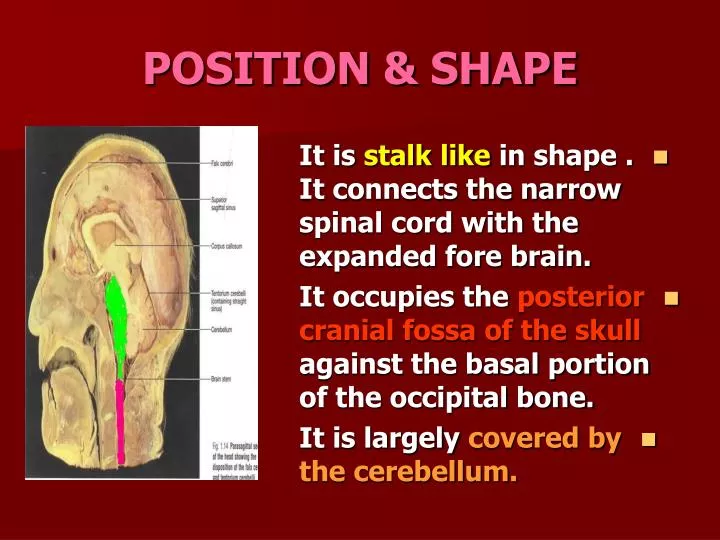
POSITION & SHAPE. It is stalk like in shape . It connects the narrow spinal cord with the expanded fore brain. It occupies the posterior cranial fossa of the skull against the basal portion of the occipital bone. It is largely covered by the cerebellum. PARTS.

E N D
POSITION & SHAPE • It is stalk like in shape . It connects the narrow spinal cord with the expanded fore brain. • It occupies the posterior cranial fossa of the skull against the basal portion of the occipital bone. • It is largely covered by the cerebellum.
PARTS • It is made up of: Medulla oblongata. • Pons. • Midbrain.
FUNCTIONS • (1)A conduit for the ascending and descending tracts connecting the spinal cord to the higher centers in the forebrain. • (2) It contains the important cranial nerve nuclei (111-x11).
FUNCTIONS • (3)It contains important reflex centers that control respiration and cardiovascular systems. • (4)It controls over the level of consciousness through the reticularformation .
CLINICAL NOMENCLATURE • It is named BULB. • The entering fibers are Cortico Bulbar. • The syndromes associated with the medulla are pseudo Bulbar palsy and Bulbar palsy.
VENTRAL ASPECT OF THE MEDULLA • It is divided by the ventral median fissure into two halves. • Each half has the following features:
PYRAMID • It is a longitudinal elevation along the side of the fissure. • The pyramids are composed of bundles of nerve fibers (Corticospinal) that originate from large nerve cells in the cerebral cortex.
PYRAMID • The pyramids taper inferiorly where the majority of the descending fibers (75-90%) cross over to the opposite side forming the Decussation of the pyramids. • This partially obscures the ventral fissure.
OLIVE It is an oval elevation lateral to the upper part of the pyramid. It is produced by the underlying Inferior Olivary Nucleus (which is connected to the cerebellum) It is important in the control of movement.
ROOTLETS OF CRANIAL NERVES • A. hypoglossal (12TH) nerve. It emerges between the pyramid and olive. • B. Glossopharyngeal,Vagus and cranial part of the Acessory (9, 10 and11TH) nerves. • They emerge in the groove between the olive and the inferior cerebellar peduncle.
PONS • It has a median groove (basilar sulcus) which lodges the basilar artery. • Its anterior surface is convex from side to side and shows many transverse Pontocerebellar fibers which are collected laterally to form the Middle Cerebellar Peduncles.
PONS • Fourcranial nerves are attached to its anterior surface. • 1. Trigeminal (5th) nerve: • It is attached to the side of the pons near its upper border by two roots . • A large sensory and a small motor. The motor root is located antero medial to the sensory root.
PONS • 2. Abducent(6th) nerve : • It is located in the groove between the lower border of the pons and the pyramid.
PONS • 3. Facial (7th) nerve : • It is found between the lower border of the pons and the inferior cerebellar peduncle. • 4. Vestibulo cochlear (8th) nerve : • It is lateral to the facial . The vestibular nerve is anterior and the cochlear nerve is posterior.
MID BRAIN • It is formed by the • Massive Basis Pedunculi(Crura Cerebri). • The crura are formed by the descending Corticobulbar and Corticospinal fibers. • The cerebral peduncles are separated by the Interpeduncular Fossa.
InterpeduncularFossa. • Boundaries: • Inferiorly :pons. • Anteriorly :optic chiasma. • Laterally: optic tracts . • Posterolaterally: cerebral peduncles.
MID BRAIN • Oculo motor (3rd) nerve : • Emerges through a groove at the medial sides of the cerebral peduncles.
DORSAL SURFACE OFTHE MEDULLA • Closed medulla • It is the continuation of the posterior surface of the spinal cord. • It is divided by the posterior median sulcus into two halves. • Each half has the following features:
LOWER MEDULLA • A.GRACILE tract: • It is a longitudinal column on both sides of the median sulcus. • It expands superiorly where it ends in the Gracile Tubercle (produced by the Gracile Nucleus).
LOWER MEDULLA • B. CUNATE tract and tubercle : • Are lateral to the gracile tract and tubercle respectively.
OPEN MEDULLA • It forms the lower third of the floor of the fourth ventricle. • It is divided by the median sulcus into two halves. • Each half has an inverted (v) shaped depression (Inferior Fovea). • It separates the motor from the sensory nuclei
OPEN MEDULLA • Medial : the hypoglossal nucleus. • Lateral : the vestibular nerve nucleus. • At the fovea : the nuclear complex of the glossopharyngeal and vagus nerves.
PONS • It is hidden by the cerebellum. • It forms the upper two thirds of the floor of the fourth ventricle. • It is widest at the pontomedullary junction. • At this point, the lateral aperture (foramen ofLuschka) is found to allow passage of CSF from the fourth ventricle to the subarachnoid space around the brain.
PONS • The posterior surface is limited laterally by the superior cerebellar peduncles and divided into symmetrical halves by the mediansulcus.Lateral to the sulcus is an elongated elevation (Medial Eminence). • Its inferior end is expanded to form the Facial Colliculus produced by the fibers of the facial nerve winding around the nucleus of the abducent nerve.
PONS • Lateral to the medial eminence is the vestibular area produced by the underlying vestibular nuclei.
MID BRAIN • It has fourColliculi(Corpora Quadregimina) which are rounded eminences that are divided into superior and inferior pairs. • Superior Colliculi: centers for visual reflexes. • Inferior Colliculi:lower auditory centers.
MID BRAIN • Trochlear nerve (the only cranial nerve that can be identified in the dorsal aspect) • It emerges in the midline immediately caudal to the inferior colliculi. • The cerebral aqueduct traverses through the length of the mid brain.
CEREBELLAR PEDUNCLES • The superior and • inferior peduncles appear on the dorsal aspect of the brain stem. • They form the lateral walls of the upper part of the floor of the 4th ventricle. • They connect the mid brain and medulla to the cerebellum respectively.
LATERAL ASPECT OF THE BRAIN STEM • The middle cerebellar pedunclecan be distinguished on the ventral, dorsal and lateral aspects of the brain stem.