Download
1 / 34
350 likes | 673 Views
#9. EBUS-TBNA for lymph nodes < 1 cm in a patient with known solitary pulmonary nodule. Describe major elements of informed consent. Describe the current evidence about staging CT/PET negative mediastinal lymphadenopathy in patients with known or suspected lung cancer.

E N D
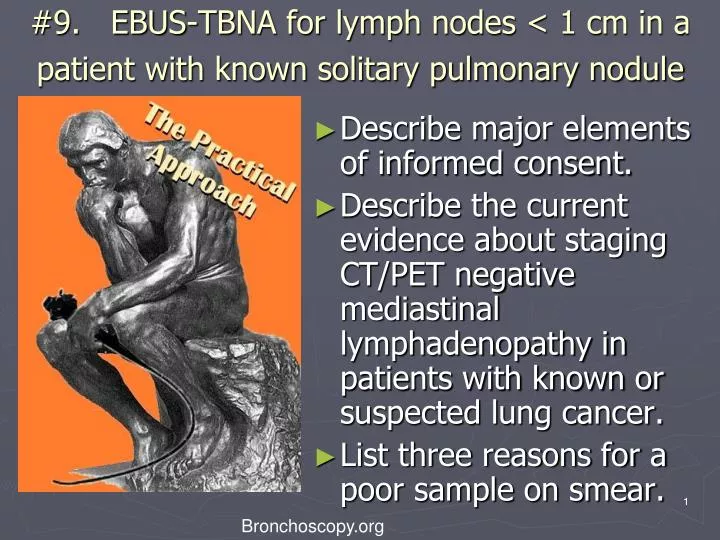
#9. EBUS-TBNA for lymph nodes < 1 cm in a patient with known solitary pulmonary nodule • Describe major elements of informed consent. • Describe the current evidence about staging CT/PET negative mediastinal lymphadenopathy in patients with known or suspected lung cancer. • List three reasons for a poor sample on smear. 1 Bronchoscopy.org
Case description(practical approach # 9) • 55 year old man with 1.5 cm solitary pulmonary nodule in the mediobasal segment of right lower lobe which was incidentally noted while he was undergoing CT of the abdomen for nephrolithiasis. • He has no risk factors for cancer. Vital signs are normal. Physical exam is unremarkable. CBC, coagulation and chemistry panel, and spirometry are normal. PPD is negative. • He is an attorney specializing in real estate law. He lives with his wife and 1 child. • He has no advanced directives. He desires all available treatment options in case he has lung cancer. Bronchoscopy.org 2
Case description(practical approach #9) • 1.5 cm RLL spn, 5.5 SUV on PET • 7 mm subcarinal lymph node on CT which is not PET avid Bronchoscopy.org 3
Initial Evaluation Procedural Strategies Techniques and Results Long term Management The Practical Approach • Examination and, functional status • Significant comorbidities • Support system • Patient preferences and expectations • Indications, contraindications, and results • Team experience • Risk-benefits analysis and therapeutic alternatives • Informed Consent • Anesthesia and peri-operative care • Techniques and instrumentation • Anatomic dangers and other risks • Results and procedure-related complications • Outcome assessment • Follow-up tests and procedures • Referrals • Quality improvement Bronchoscopy.org 4
Initial Evaluations • Exam • ECOG performance status 0 • Comorbidities • Nephrolithiasis • Support system • Wife and teenage child all healthy and actively involved with patient’s care. • Patient preferences • Desires all available active treatment options. Bronchoscopy.org 5
Procedural Strategies • Indications • Invasive lymph node staging in a patient with radiographically (CT and PET) normal mediastinum • PET scan and Integrated PET/CT have demonstrated a high NPV for mediastinal nodal disease with NPV of ~90%* • 5.6% of patients with radiographically stage 1 disease were found to have N2 disease** • Risk factors and incidence of occult N2 mets include*** • larger tumor size (≥6.0 cm) – 57% prevalence • central location- 21.6% prevalence • high PET SUV (≥4.0)- 10.5% prevalence • adeno ca cell type- 9.0% prevalence NPV PET vs. EBUS in subgroup with AdenoCA 77.8 % and 96.7% respectively (p= 0.011) *Eur Respir J 2009; 33: 201–212 **J Thorac Cardiovasc Surg 2006;131:822-829 ***Ann Thorac Surg 2007;84:177-181 6 Bronchoscopy.org
Procedural Strategies *Ann Thorac Surg 2007;84:177-181 **J Thorac Cardiovasc Surg 2006;131:822-829 *** Eur J Cardiothorac Surg. 2007 Jul;32(1):1-8 • Indications • Should this patient undergo lymph node staging before or after needle aspiration biopsy of his peripheral nodule? • Should he undergo invasive lymph node staging? • Invasive staging should be reserved for patients with 1 or more risk factors for occult N2 disease* ** *** • Routine use of invasive lymph node staging for patients with clinical stage 1 non small cell lung ca and no risk factors for occult N2 disease is not cost effective** Bronchoscopy.org 7
Procedural Strategies *Chest 2008;133;887-891 **Eur Respir J 2009; 33: 1156–1164 • Contraindications: • None • Expected Results: • EBUS-TBNA demonstrated 89% sensitivity, 100% specificity, 98.9% NPV for 5-10 mm radiographically normal nodes* • Operator and team experience: well experienced team • Risks-benefits: • Safe procedure with no serious complications reported in the literature. • Agitation, cough, and presence of blood at puncture site have been reported as infrequent complications.** • Benefits: highly accurate and safe procedure to obtain tissue for staging. Same day procedure. Bronchoscopy.org 8
Procedural Strategies *AM J RESPIR CRIT CARE MED 2000;161:601–607 **Thorax 2005;60:949–955 ***Chest 2007;132;202-220 **** JAMA. 2008;299(5):540-546 *****J Bronchol 2008;15:17–20 ****** Lung Cancer 64 (2009) 127–128 • Diagnostic alternatives: • Transbronchial needle aspiration • Sensitivity 65-89% for subcarinal node location > 1cm* • Sensitivity 39% if median prevalence of disease is 34%** • High false negative rate 28%*** requires negative results to be followed with another invasive staging procedure • Histology needles provide increased diagnostic yield except when lesions are <1 cm* • In a head to head comparison sensitivity/NPV was 36%/78% and 69%/88% for TBNA vs EBUS-TBNA respectively**** • A therapeutic strategy using both TBNA and EBUS TBNA**** or TBNA and EUS-FNA *****has been proposed as the most cost effective strategy for staging the mediastinum BI #. Practical Approach Title 9
Procedural Strategies *JAMA. 2008;299(5):540-546 **Chest 2007;132;178S-201S ***Eur J Cardiothorac Surg. 2007 Jul;32(1):1-8 ****Chest 2007;132;202-220 • Diagnostic alternatives: • Percutaneous needle aspiration of nodule • Endoscopic ultrasound in a head to head comparison* sensitivity and negative predictive value (69% and 89% respectively) was identical to EBUS for accessible nodes • Mediastinoscopy gold standard. • Mediastinoscopy is invasive staging procedure of choice in radiographically negative mediastinum** *** • 42% sensitivity in clinical stage 1 **** • Difficult to access level 7 nodes • VATS most invasive of alternatives. Only provides access to ipsilateral nodes. 75% sensitivity****. Benefits include definitive lobar resection at same time if node negative. Bronchoscopy.org 10
Procedural Strategies Risks-Benefits Cost effectiveness- no formal evaluations have been published In 2 separate decision-analytic models, both (EUS-FNA + EBUS-FNA) and (conventional TBNA + EBUS-FNA) were more cost-effective approaches than mediastinoscopy for staging patients with NSCLC and abnormal mediastinal lymph nodes on non-invasive imaging* ** A strategy adding EUS-FNA to a conventional lung ca staging approach (mediastinoscopy thoracotomy) reduced costs by 40% per patient*** May actually increase health care costs if done in low volume centers by less experienced operators**** ***** Start up costs Cost of equipment ~100K******and training Physician reimbursement ~$280; facility reimbursement $257****** *Gastrointestinal Endoscopy 69, No. 2, Supp 1, 2009, S260 **J Bronchol 2008;15:17–20 ***Thorax 2004;59;596-601 ****Lung Cancer 64 (2009) 127–128 *****J Bronchol 2008; 15:127-128 ****** Southern Medical Journal 2008;101,No5;534-38 Bronchoscopy.org
Procedural Strategies Drawing from Herth FJ et al.. Chest 2003;123:604 –7 EBUS image from patient. • Informed consent: • There were no barriers to learning identified. Patient has good insight into his disease and realistic expectations. BI #. Practical Approach Title 12
Procedural techniques and resultsAnesthesia and perioperative care Conscious sedation May be performed in bronchoscopy suite Cost savings compared to general anesthesia. Visualization and biopsy of smaller nodes technically more difficult than with general anesthesia. General anesthesia with LMA Better visualization of higher nodes compared with ET tube May be performed in bronchoscopy suite May not be appropriate in severe obesity or severe untreated GERD General anesthesia with ET tube Usually performed in OR EBUS scope directed more centrally in airway which may make biopsies more difficult 13 Bronchoscopy.org Chest 2008;134;1350-1351
Procedural Techniques and Results • Instrumentation • EBUS scope- direct real time US imaging with curved array ultrasound transducer incorporated in distal end of bronchoscope • Ultrasound processor • Adjustable gain and depth • B mode and Doppler capabilities • Needle • 22 gauge acrogenic needle with stylet • Needle guide system locks to scope • Lockable needle and sheath • Precise needle projection up to 4 cm Bronchoscopy.org 14
Procedural Techniques and Results Chest 2004;126;122-128 **Eur Respir J 2002; 19:356–373 ***Eur Respir J 2009; 33:935-938 • Anatomic dangers and other risks • Major blood vessels- azygous, PA, aorta, SVC and Left atrium • Risk of canulating major vessel significantly reduced with real time B mode and Doppler mode imaging • “Minor” oozing of blood at puncture site was reported in 1 study there have been no reports of major bleeding* • Pneumothorax and pneumomediastinum** • Have been reported with blind TBNA but no reports in literature with EBUS guided FNA. • A case of bacterial pericardial effusion and nodal infection have recently been reported as complications following EBUS with full needle extension***. Bronchoscopy.org 15
Procedural Techniques and Results Aspirate cytology Adequate/representative: in presence of frankly malignant cells, lymphocytes, lymphoid tissue, or clusters of anthracotic pigment-laden macrophages* Inadequate/nonrepresentative: if there are no cellular components, scant lymphocytes (defined as <40 per HPF) blood only, or cartilage or bronchial epithelial cells only* ** A quantitative cut off value of at least 30% of cellularity composed of lymphocytes has been arbitrarily proposed by some experts*** Higher yield may be obtained by obtaining aspirates from the periphery of nodes**** *Am J Clin Pathol 2008;130:434-443 **Chest 2008;134;368-374; ***Chest 2004;126;1005-1006 ****Techniques in GI Endoscopy, Vol 2, No 3, 2000: pp 136-141
Procedural Techniques and Results • Number of aspirates* if ROSE not utilized • Best yield with 3 aspirates per station (see table) • Two aspirations per LN station can be acceptable when at least one tissue core specimen is obtained by the first two aspirations • Sensitivity 91.7%, NPV 96.0%, and accuracy 97.2% • If operator believes targeting is inadequate or insufficient another aspirate should be performed 17 Bronchoscopy.org * Chest 2008;134;368-374;
Maximum results after 3 aspirates Rapid On Site Cytology is standard of care to assure greater yield and better patient care Chest 2008;134;368-374 Bronchoscopy.org
Procedural Techniques and Results • Results and procedure-related complications • Due to the absence of history suggestive of noncancer diagnosis and high SUV on PET the level 7 node was sampled with EBUS under general anesthesia using a 9.0 ET tube. • The cytology was diagnostic for non small cell carcinoma (adenocarcinoma). • There were no complications. Bronchoscopy.org 19
Long-term Management Plan • Outcome assessment • Patient was referred for multidisciplinary evaluation to include cardiothoracic surgery, oncology, and radiation oncology for potential trial enrollment for neoadjuvant treatment of stage IIIA adenocarcinoma of the lung.* • Follow-up tests and procedures • Patient will follow up in 1 month to ensure he has been evaluated by all the above specialties. • Referrals • See above. • Quality improvement • The occult N2 metastasis was identified prior to the patient undergoing VATS or open thoracotomy for a non resectable tumor. • 5 year survival for IIIA non-small cell lung ca is 23%. *Chest 2007;132;243S-265S Bronchoscopy.org 20
Q 1: Describe the major elements of informed consent Bronchoscopy.org
The concept of Informed Consent • Protects the patient by providing them with complete information on which to make an informed decision. • Protects the health care provider from liability provided the procedure is properly executed according to the prevailing standards of care in the community and without negligence. • Gives the health care providers an opportunity to consider and re-consider the diagnostic and therapeutic strategies being proposed • Allows for a discussion of possible risks and benefits and to prepare for procedure-related events. Bronchoscopy.org
The requirements of Informed Consent From a legal standpoint, consent for a medical procedure must be both informed and effective. To be informed, a patient must be given information about the procedure relevant to their individual situation. To be effective, the person undergoing the procedure should be able to demonstrate, in his or her own words, their understanding of the procedure or treatment. Bronchoscopy.org
American Medical Association: Informed consent is a process which should disclose and discuss: • The patient’s diagnosis and concerning clinical issues. • The nature and purpose of the proposed procedure • The risks and benefits of the proposed procedure. • Alternative regardless of cost or coverage by health insurance. • Potential risks and benefits from choosing the alternatives. • The risks and benefits of not receiving or undergoing treatment or procedures. Bronchoscopy.org
Q 2: Describe the current debate about staging CT/PET negative mediastinal lymphadenopathy in patients with known or suspected lung cancer. Bronchoscopy.org
Q 2: Describe debate about staging CT/PET negative mediastinal lymphadenopathy • Question • Should these patients undergo invasive lymph node staging? • Invasive staging should be reserved for patients with 1 or more risk factors for occult N2 disease. • Routine use of invasive lymph node staging for patients with clinical stage 1 non small cell lung ca and no risk factors for occult N2 disease is not cost effective. BI #. Practical Approach Title 26
Q 2: Describe current evidence about staging CT/PET negative mediastinal lymphadenopathy • Facts • PET scan and Integrated PET/CT have demonstrated a high NPV for mediastinal nodal disease with NPV of ~90%. • 5.6% of patients with radiographically stage 1 disease have N2 disease. • Risk factors and incidence of occult N2 disease: • larger tumor size (≥6.0 cm) – 57% prevalence • central location- 21.6% prevalence • high PET SUV (≥4.0)- 10.5% prevalence • adeno ca cell type- 9.0% prevalence Bronchoscopy.org 27
EBUS is warranted for staging patients with PET negative, normal mediastinum • Patients highly suspicious for NSCLC with CT scans showing no enlarged lymph nodes (no node > 1 cm) and a negative PET finding of the mediastinum underwent EBUS-TBNA. • Identifiable lymph nodes at locations 2r, 2L, 4r, 4L, 7, 10r, 10L, 11r, and 11L were aspirated. • All patients underwent subsequent surgical staging. Diagnoses based on aspiration results were compared with those based on surgical results. • One hundred patients (mean age, 52.4 years; 59 men) were included. After surgery, 97 patients (mean age, 52.9 years; 57 men) had NSCLC confirmed and were included in the analysis. Herth F et al. CHEST 2008; 133:887–891 Bronchoscopy.org
In this group, 156 lymph nodes ranging 5 to 10 mm in size were detected and sampled. • Malignancy was detected in nine patients but missed in one patient. • Mean diameter of the punctured lymph nodes was 7.9 mm. • The sensitivity of EBUS-TBNA for detecting malignancy was 89%, specificity was 100%, and the negative predictive value was 98.9%. No complications occurred. • EBUS-TBNA can be used to accurately sample and stage patients with clinical stage 1 lung cancer and no evidence of mediastinal involvement on CT and PET. • Potentially operable patients with no signs of mediastinal involvement may benefit from presurgical staging with EBUS-TBNA. Bronchoscopy.org Herth F et al. CHEST 2008; 133:887–891
Bronchoscopy.org Herth F et al. CHEST 2008; 133:887–891
Q3: List three reasons for a poor sample on smear Bronchoscopy.org
Diagnostic BronchologySampling and Smearing: tips and tricks. • Avoid: • Sample coagulation : delay in processing the sample may completely hinder the process of smearing on the slide because of its coagulation, even when the blood content of the sample is not too much. Sample coagulation may happens in the needle lumen or on the slide surface. • Air drying artefacts : if Papanicolaou (fig. A&B) stain is required, the smear must be still wet when fixative is applied, otherwise staining artefacts are the rule. Conversely, with Diff-Quick stain, the quantity of sample fluid must be just enough to smear, enabling the sample to dry as soon as possible. • Air bubbles : their presence in the material put on the slide causes uneven smear distribution. This problem happens mainly with needle aspiration technique. • Thick smear : the correct technique of smearing gives as final result distribution of cells in monolayer on slide surface; cells overlapping makes the diagnostic process difficult or impossible because it is impossible to evaluate precisely cellular details. To avoid this problem smear only a small drop of sample on the slide surface. • Crushed smear : is the result of excessive pressure applied to smear the sample; this may completely destroy diagnostic material. Don’t worry, this may take some time to learn ! Needle biopsy of peripheral lung nodule (adenocarcinoma). Fig. A and B come from the same sampling and staining process. The slide of fig.A was correctly smeared and fixed while wet, giving precise color profile (pale blue); the slide of fig. B was correctly smeared but fixed when completely dried giving wrong color profile (pinkish): same neoplastic cells, same staining procedure, different color profile ! A B Bronchoscopy.org From G. Marciano, with permission
Bronchoscopy International: Practical Approach, an Electronic On-Line Multimedia Slide Presentation. http://www.Bronchoscopy.org/PracticalApproach/htm. Published 2009 (Please add “Date Accessed”). All efforts are made by Bronchoscopy International to maintain currency of online information. All published multimedia slide shows, streaming videos, and essays can be cited for reference as: Thank you Bronchoscopy.org 33
Prepared with the assistance of Steven Escobar M.D., Balboa Navy Medical Center, San Diego, CA www.bronchoscopy.org Bronchoscopy.org 34


![Respiratory Physiology [the Ins and Outs]](https://cdn2.slideserve.com/4330665/respiratory-physiology-the-ins-and-outs-dt.jpg)