Download
1 / 41
520 likes | 5.48k Views
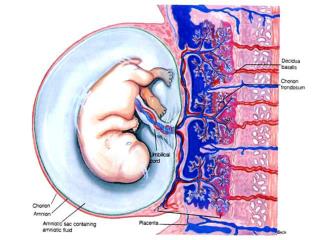
Ultrasound of Placental Abnormalities. Guy Steinberg, MD March 5, 2011. Initial examination. 5-7 Weeks - placenta is a diffusely echogenic ring. By 8-13 Weeks - focal chorionic thickening and determination of placental site, umbilical cord inserts in center of the chorionic frondosum.

E N D
1. Ultrasound of Placental Abnormalities Guy Steinberg, MD
March 5, 2011
2. Initial examination 5-7 Weeks - placenta is a diffusely echogenic ring.
By 8-13 Weeks - focal chorionic thickening and determination of placental site, umbilical cord inserts in center of the chorionic frondosum.
R/o implantation on fibroid and succinturiate lobes.
Implantation in areas with less blood supply, such as a myoma or a uterine septum, can be associated with later complications such as spontaneous miscarriage, IUGR, preterm labor, placental abruption and PPH.
3. Initial examination
Second trimester placenta - More heterogeneous, sonolucencies are common.
Prominent sonolucencies in the second trimester usually result in a normal outcome, and is more common in women with elevated MSAFP or first trimester bleeding. 2-18% of placentas have sonolucencies at 15-34 W.
Usually are venous lakes with minimal blood flow , and eventually have fibrin deposition. May be more than 1 cm in diameter, have swirling blood flow, and may change in shape during the exam.
More often seen in thick placentas - placenta > 3 cm is 6X more likely to have sonolucencies. If extensive (>3), large (>2cm), or if present before 20-25 W f/u and look for signs of placental insufficiency - IUGR, oligo, doppler.
�Swiss cheese� placental variant: patchy placenta from diffuse fibrin deposition, many sonolucencies, placentomegaly, r/o gestational trophoblastic neoplasia. DD: chorioangioma, abruption.
5. Placenta Previa Low lying placenta (<2cm), marginal previa (edge at internal os) and complete previa (internal os completely covered by placental tissue). Epidemiology: 5% incidence at 15-16 W, 0.5% at term.
Risk factors: AMA, multiparity, smoking, cocaine use, prior previa, previous CS, uterine transcavitary surgery as well as prior vigorous sharp curettage.
Placenta previa is associated with accreta/perceta spectrum: abnormal placenta may grow through endometrium. 5% of placenta previa have associated accreta/perceta. Higher risk if if prior CS and anterior PP, increased risk with increasing prior CS. 67% risk if previa and >4 CS.
Look for hypoechoic line between bladder and placenta.
6. Placenta Previa Placental migration is the displacement of the placenta from a low or previa implantation to a higher position in the uterus.
Likely involves peripheral degeneration secondary to decreased vascularization with preferential growth in areas assumed to be optimally perfused.
May involve more rapid growth of the lower uterine segment, as compared to the rest of the uterine body.
Trophotropism: atrophy is areas of low blood supply (LUS) and growth in areas of better blood supply. Placental �migration� is about 5mm/wk.
8. Vasa Previa Fetal vessels run through the membranes, unprotected by placental tissue or Wharton's jelly, below the fetal presenting part and close to the internal cervical os.
It should particularly be suspected when the placental edge covers the os in mid-pregnancy but recedes later on. Occurs in 1/2500 deliveries.
Risk factors include multiple pregnancy, pregnancy resulting from IVF, presence of velamentous insertion of the umbilical cord, succenturiate or accessory placental lobes.
Velamentous insertion of the cord refers to vessels traveling through the fetal membranes before entering the actual placenta, as opposed to the normal insertion of the cord directly into the placenta.
This has to be differentiated from marginal insertion of the cord, when the insertion is at the placental edge, rather than more centrally.
9. Vasa Previa It should be distinguished from funic presentation, when loops of a normally implanted cord are preceding the presenting part at the internal os.
With funic presentation, manual displacement of the presenting part and manipulation of the lower segment, generally demonstrate movement of the loops of cord.
US Findings: More common: vasa previa from succinturate lobe, or vasa previa from velamentous cord insertion.
TVS + doppler required for Dx: color doppler shows crossing vessels, pulsed doppler shows fetal vascularity.
Most commonly an incidental finding with succinturate lobe. 60-80% fetal mortality if diagnosis missed.
11. Placenta accreta Abnormal adherence of the placenta to the uterus, due to an absence or deficiency of Nitabuch's layer or the spongious layer of the decidua.
Invasion to the myometrium is defined as placenta increta, and invasion through the myometrium and serosa is called placenta percreta.
The incidence of placenta accreta is rising, primarily because of the rise in CS deliveries. The risks increase with the number of previous CS: 4-fold after one previous CS, up to 11.3 RR increase after two.
Patients with a previous CS and previa implantation of the placenta are 5X more likely to have placenta accreta. Another etiological factor is local tissue alteration, secondary to curettage.
12. Placenta accreta Prenatal diagnosis of placenta accreta by imaging should be based on the irregularly shaped placental lacunae signs (swiss cheese appearance) representing vascular spaces rather than only the loss of the normally obvious retroplacental clear space.
Other signs include thinning of the myometrium overlying the placenta, protrusion of the placenta into the bladder, increased vascularity of the uterine serosa bladder interface, and turbulent flow through the lacunae using Doppler studies.
14. Placental abruption This is a leading cause of vaginal bleeding in the latter half of pregnancy, complicating about 0.5-1% of pregnancies.
Risk factors are defined and consist of abruption in a previous pregnancy, maternal hypertension, AMA, smoking, cocaine use, trauma and some uterine anomalies.
The diagnosis of abruption is a clinical one, and ultrasonography is of limited value with a sensitivity of no more than 50%.
On ultrasound one might see placental edge separation, subchorionic and retroplacental hematomas.
In the acute phase, the area of detachment may appear hyperechoic, becoming hypoechoic after a few days.
15. Placental bed infarction Also known as intervillous thrombosis, this is a vascular lesion caused by thrombosis in the villous spaces (ischemic necrosis).
Round, mostly anechoic, intraplacental, measuring up to several centimeters. It is common and, unless massive, of minimal clinical significance.
Large infarcts or those occurring in early pregnancy may reflect underlying maternal vascular disease.
It is different from maternal floor infarction (not a true infarct), a recurrent lesion of unknown etiology, in which fibrin is deposited throughout the placenta, leading to necrosis of villi, and often accompanied by fetal demise.
16. Hematomas These appear as crescent-shaped, sonolucent fluid. These can be on the fetal (retrochorionic, subchorionic) or maternal (retroplacental) side.
Retrochorionic hematomas are lesions involving maternal blood separating the chorionic membrane from the villous chorion.
Minimal clinical significance, unless + vaginal bleeding or uterine contractions when they can be markers of later complications, such as abortion, IUGR, PTL, placental abruption and fetal distress.
Commonly confused with chorioangiomas.
17. Hematomas Subamniotic hematomas - between the amniotic and chorionic membrane and result from rupture of chorionic vessels, near the cord insertion.
Clinically more significant, retroplacental hematomas are typically located between the placental basal plate and the uterine wall.
They usually bulge towards the fetal side and result from bleeding from the spiral arteries.
Associated with perinatal complications, secondary to the presence of blood. Management consists of close observation.
19. Placental thickness/size (volume) Placental size has been shown to be an important factor in abnormal fetal growth. In daily clinical practice, it is not routine to measure placental thickness.
During examination of the placenta, thickness is grossly evaluated and, if appearing normal to the eye, is not further delineated.
Only if suspicious, will the maximal vertical thickness be measured.
Increased thickness is non-specific and can be found with maternal diabetes, maternal anemia, abnormalities of fetal growth, fetal hydrops, and TORCH infections.
20. Placental thickness/size (volume) Placenta thickness increases with gestation: 10mm at 10W, 20 mm at 20 W, 30mm at 30 W - >40 mm considered placentomegaly. Mature weight 250-550 gr, 16-20 cm in diameter.
DD of placental thickening/placentomegaly: placental insufficiency, diabetes, maternal or fetal anemia, hydrops, intrauterine infection, Beckwith-Wiedemann syndrome, hydatiform mole, chromosomal abnormalities, placental mosaicism.
A thickened placenta is usually nonspecific and associated with a normal outcome.
A very thickened placenta n early pregnancy increases risk of IUGR and placental insufficiency.
22. Circumvallate and circummarginal placenta When the chorionic plate is smaller than the basal plate of the placenta, the placental periphery is not covered, leading to some of the placenta to be extrachorial.
Circumvallate placenta (1-2%) is caused when the fetal surface presents a central depression surrounded by a thickened ring, composed of a double fold of amnion and chorion with degenerated decidua and fibrin in between.
This creates an irregular edge with uplifted margins or placental sheet or shelf.
Linked to placental abruption, fetal hemorrhage and accordingly to adverse perinatal outcome, including increased fetal morbidity and mortality.
When the ring is flat, rather than and lacks central depression, it is called circummarginal placenta (15-20%), a condition with questionable clinical significance.
24. Accessory lobe: succenturiate placenta Succenturiate placenta : one or more small accessory lobes that develop at a distance from the main placenta.
A smaller version of a bilobate placenta which occurs when two approximately equal lobes of placenta are found.
The�recognition of this condition is important since intramembranous vessels connecting the main placenta with the succenturiate placenta may rupture during labor with rupture of the membranes (vasa previa) and fetal death may ensue.
Retention of an accessory lobe may lead to PPH.
25. Placental grading Premature aging (grade 3 before 37 W) is associated with perinatal complications, such as IUGR and fetal distress.
Grade 3, early in pregnancy is associated with IUGR and/or smoking, chronic hypertension, SLE, diabetes and other vascular disease.
Its value in low-risk pregnancies is limited, but some studies have suggested that ultrasound detection of a grade 3 placenta at 36 W might help to identify a high risk pregnancy, in terms of subsequent development of gestational hypertension and IUGR.
27. Placental calcifications Increases with gestational age and is recognized as a normal part of maturation and aging. Also described in several infectious processes.
Numerous small, echogenic nodules on the fetal surface may be consistent with amnion nodosum or squamous metaplasia.
Amnion nodosum is a localized, diffuse accumulation of amorphous material (vernix, desquamated fetal skin cells, epithelial cells, and hair) producing small nodules on the placental surface. It is typically associated with fetal renal agenesis, oligohydramnios and poor fetal outcome.
Squamous metaplasia is similar, localized more around the umbilical cord insertion. It has no clinical significance, and has been not been described by US.
28. Gestational trophoblastic disease (GTD) Proliferative disorders of the placental trophoblast -benign (80%) and malignant (20%). Increased �-hCG secretion characterizes this condition which is highly curable even in the presence of metastases.
The major risk factors for gestational trophoblastic disease are Asian ethnicity, AMA, and a past history of GTD.
Common US presentations include enlarged uterus and absence of fetal parts and a �snowstorm� appearance without embryonic structure.
Maternal lakes resulting from stasis of blood between the molar villi are common as well. Large ovarian theca lutein cysts, secondary to the elevated �-hCG levels may also occur.
Complete moles: secondary to sperm fertilizing ovum with inactive nucleus, haploid sperm duplicates to diploid - 47XY karyotype.
29. Gestational trophoblastic disease (GTD) The partial mole: focal trophoblastic hyperplasia with villous hydrops together with identifiable fetal tissue.
Fertilization of a normal oocyte by more than a single sperm or a diploic sperm or fertilization of a diploic oocyte by a normal haploid sperm, all resulting in a conceptus with 3 sets of chromosomes (69XXX or 69XXY).
> 90% of triploid fetuses show severe, symmetrical IUGR and multiple anomalies, commonly accompanied by oligohydramnios and abnormal placental Doppler indices.
The typical placental molar features are not always present in triploid partial moles and are less likely to be apparent on US in early pregnancy.
Malignant trophoblastic disease occurs in 15-20% of patients with hydatidiform mole but rarely in partial mole - described as irregular echogenic lesions within the myometrium, often surrounded by echofree areas, corresponding to local hemorrhage.
32. Chorioangioma Chorioangioma : most common benign nontrophoblastic placental neoplasm - a hypervascular cystic lesion, single or multiple, with arterial and venous flow that produces color signals.
US: well-circumscribed, rounded, hypoechoic lesion next to the chorionic surface, often close to the cord insertion.
Small lesions are often present when examining the placentas of completely asymptomatic, normal pregnancies.
Larger lesions (>5�cm) are rare, can be associated with non-immune hydrops, IUGR, neonatal cardiomegaly, anemia and thrombocytopenia) and mortality, as well as maternal morbidity such as polyhydramnios and pre-eclampsia.
Color Doppler US is useful in the early diagnosis and differentiation from hematomas as well as evaluation of response to treatment.
34. Placental cyst Placental cyst is a rare, mostly benign, subchorionic lesion, filled with mucoid material.
It is a single finding in most cases, but, occasionally, multiple cysts are present.
Only if large (>4.5�cm) have they been associated with fetal complications, such as IUGR or fetal demise.
36. Placenta in multiple gestations Monochorionic twins are at risk for type-specific perinatal complications, for example, twin twin transfusion syndrome (TTTS).
There is a higher frequency of velamentous and marginal cord insertion in twins, potentially leading to IUGR as a result of unequal sharing of placental tissue.
It is estimated that 95% of MC twin placentas contain interfetal vascular connections.
These connections may be indirect artery-to-vein (A-V), found in up to 80% cases (within the placental villi), direct artery-to-artery (A-A), in 75% (at the fetal surface), or direct vein-to-vein (V-V) in 20% (at the fetal surface).
37. Placenta in multiple gestations The A-V connections are responsible for TTTS, while A-A may protect against it but lead to twin reversed arterial perfusion (TRAP), together with V-V connections.
In TRAP sequence, one twin (pump twin) forces blood to the entire placenta and the acardiac twin who acts as an additional cotyledon but without reoxygenating that amount of blood that passed through it.
Sluggish flow increases risk of thrombosis and thromboembolic phenomena in the pump twin.
Cord entanglement is another risk specific to monoamniotic twins.
38. Placenta in multiple gestations Membranes in a monochorionic pregnancy has been described with the T sign: membrane inserts the placenta at a 90 degree angle.
The presence of a thick (>2�mm) dividing membrane, from 10 - 14 weeks, supports the diagnosis of dichorionicity.
The presence of the lambda sign or T sign in the presence of a single placenta, in the first trimester, is the most reliable indicator of chorionicity.
This analysis of the membranes should be performed in higher rate multiple gestations in a similar fashion.
41. Thank you