Download
1 / 21
210 likes | 303 Views
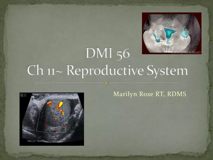
DMI 56 Ch 11~ Reproductive System. Marilyn Rose RT, RDMS. outline. Infectious diseases of both genders Male reproductive system Physiology Benign prostatic hypertrophy Carcinoma of the prostate gland- staging Undescended testis- cryptorchidism Testicular torsion/ epididymitis

E N D
DMI 56Ch 11~ Reproductive System Marilyn Rose RT, RDMS
outline • Infectious diseases of both genders • Male reproductive system • Physiology • Benign prostatic hypertrophy • Carcinoma of the prostate gland- staging • Undescended testis- cryptorchidism • Testicular torsion/ epididymitis • Testicular tumors • Female reproductive system • Physiology • PID • Cysts and tumors • Ovarian cyst/ tumor • Dermoid cyst • Uterine fibroids • Endometrial carcinoma • Endometriosis • Carcinoma of the cervix • Breast lesions- cancer/ benign • Pregnancy- ectopic/ trophoblastic • Female infertility
Syphilis • Chronic/ Sexually transmitted / systemic infection • Spirochete Treponemapallidum • Baby- congenital syphilis • Primary- chancre- on genitals • Secondary- untreated stage one- nonitching rash- any part of the body- still infectious • Up to now cured with antibiotics… • Tertiary- untreated secondary…radiographic abnormalities • Now incurable…. • Common in young black male population
Radiographic syphilis • Cardiovascular • ascending AO- aneurysmal and linear ca++ • Skeletal system • Findings of chronic osteomyelitis- long bones/ skull • Neuropathic joint disease (Charcot’s joint) • Cerebral cortex- cause mental d/o, deafness, blindness
Gonorrhea • Bacterial infection- one of the most common (men) • Acute urethritis with copious discharge of pus • Women can be asymptomatic • If untreated- chronic inflammation spreads upward • Fibrosis- urethral stricture in men • PID in women- fibrous scarring of fallopian tubes • Radiographic- septic arthritis, PID- use US to see abnormal fallopian tubes or ectopic pregnancy • Responds to antibiotics
Male reproductive system • Formation of sperm • Begins about 13 and continues for life • FSH from pituitary- produce spermatozoa • Male testes secrete – testosterone • Stimulates accessory sex organ development and male sexual behavior • Body hair, deep voice, growth of skeletal muscles • Maturation of sperm in epididymis- vas deferens- joins seminal vesicle to form ejaculatory duct • Vasectomy= severing the vas deferens • Prostate gland is located below the bladder- surrounds the urethra- secretes alkaline fluid that promotes motility of the sperm • Male fertility is related to the number of sperm and their size, shape and motility • Sterility is <50 million per ml of semen- yet it only take one!!
Benign prostatic hyperplasia -BPH • Enlargement of prostate gland • Common in men over 60 yrs • Major effect-inability to empty bladder completely-\ • Partial urinary tract obstruction • Hydronephrosis • Transrectal US- gland enlargement ( distinguish from prostate ca) • TURP- (transuretheral resection) removal of prostate can relieve the obstruction
Carcinoma of prostate • 2nd most common malignancy in men (> black men) • >40% increase with advancing age • Tumor can be slow growing or aggressive with mets • Peripheral zone – 70% • Best detected by palpatation- hard nodular, irregular mass on rectal exam • >PSA serum= abnormality though not specific for malignancy • Radiographic= impresses floor of bladder, with irregular shape (smooth in BPH), bladder neck obstructions, infiltration of the trigone or invasive obstructions of ureters above the bladders can obstruct the upper urinary tract. • Transrectal US- once preferred- areas of low echogenicity in the prostate- 40% isoechoic to normal prostate tissue, so not conclusive with US • MRI is superb for accurate staging for pelvic neoplasms
Undescended testis- cryptorchidism • Towards the end of gestation the testes migrate from the abdominal cavity to the scrotal sac though the inguinal canal • Premature males- can cause infertility • If one of the testicles cannot be palpatated- determine whether this represents absence or ectopic position • Rate of malignancy 40% > in undescended testicle • Radiographic: • US used to screen- and especilly locate testis in inguinal canal. • Not as sensitive for ectopic testicles in pelvis or abdomen. Only if US cannot locate the testicle then MRI or CT is indicated.
Testicular torsion/ epididymitis • Twisting of the gonad on its pedicle • Compromise of circulation • sudden onset of severe scrotal pain • Color doppler US is helpful to evaluate for blood flow- • Torsion < flow to arterial perfusion or absent • Epididymitis > flow • Immediate surgery within 5-6 hours is necessary in torsion to preserve the testicle • Epididymitis- antibiotics and rest
Testicular tumor • Most common neoplasm- men 20-35 years of age. • testicular tumors are malignant • metastasize to the lymphatics • Two major types • Seminoma 45%- seminiferous tubules • Germ cell tumors 55% (teratoma)- primitive germ cells • US is best • circumscribed mass with either > or < echo • Seminoma- uniform hypo w/out ca++ or cyst • Teratoma- inhomogeneous with cystic and solid and ca++
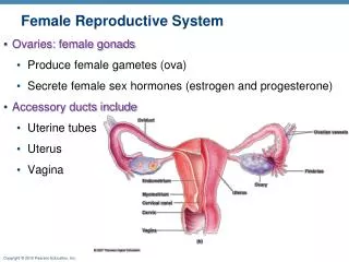
Female reproductive system • The ovaries- produce ova and secrete female hormones • Menarche- onset of menstruation • 1st day graafianfolliclees/ova develop- secrete estrogen • One migrates to surface /ruptures the egg (ovulation) • For 7-8 days a corpus luteum continues to grow where the egg was released- it secretes progesterone • No egg fertilization- it becomes corpus albicans- scar • Fertilization- corpus luteum remains intact to keep the pregnancy viable.
Female reproduction contd. • Female cycle is controlled by the pituitary • FSH- graafian, ova, estrogen • LH – rupture , progesterone • Fallopian tubes serve as ovary ducts- where fertilization occurs • In the next few days the embryo reaches the endometrium- UT • If the embryo implants in the fallopian tube- ectopic pregnancy • In 10 days a placenta develops- blood crosses the fetal membrane or chorion • Proliferative phase- between the end of menses/ ovulation • Secretoryphasee- occurs between ovulation and onset of menses • Menopause- when reproductive years terminate / menstrual periods cease- early 50’s
PID • Inflammation of the pelvic reproductive organs • Result of venereal disease (gonorrhea) • Peak 20-24 years • Unsterile abortion, delivery, multiple sexual partners or a complication from IUD • Not treated? Spread infection to fallopian tubes and cause scar/ fibrous adhesions • Outer ends stay open? peritonitis or pelvic abscess • Outer ends close? Pyosalpinx and heal to form a hydrosalpinx • Obstruction of fallopian tubes- can cause infertility or ectopic pregnancy • US is best to demonstrate fluid filled fallopian tubes • Abscess demonstrates irregular or “shaggy “ wall fluid collection with debris and often pain, tenderness and hypervascular color Doppler • Can evaluate the fallopian tubes for patency with a hysterosalpingogram
Ovarian Cysts and tumors • Physiological cysts are most common in female infants and in women of childbearing age • Follicular cyst • Corpus luteum cyst • PCOS is most common genital disorder in women • Most common malignancies of ovaries • metastatic tumors from breast, colon and stomach- bilat • Radiographic: • US – round, anechoic fluid filled “sacs” on one/both ovaries • PCOS- 10 or more cysts per ovary- peripherally “string of pearls” • Primary cystadenocarcinoma- ca++ in a fluid filled sac/ septa and solid components • can be difficult to distinguish between the benign cystadenoma… • the addition of ascites - more convincing for a malignant process • Dermoid- most common germ cell tumor • with skin, hair, teeth, and fat- no clinical significance- • an echogenic mass of the ovary- should be removed so it does not turn into a malignancy • MRI is most definitive to characterize the ovarian masses
Uterine fibroids (myomas) • Very common benign smooth muscle tumors • Multiple and vary in size • Growth is stimulated by estrogen • Develop only during reproductive years • Shrink after menopause • Common symptoms= • abnormal bleeding between periods • excessively heavy flow • Large tumors can compress surrounding organs • Can interfere with delivery if by the cervix • Radiographic- • Most common calcified lesion of female genital tract • A whirl appearance with popcorn calcification or a thin calcified wall • A myomectomy is sometimes done …new advances are multilaser and myolysis – electrical coagulation or uterine artery embolization
Endometrial carcinoma • Adenocarcinoma of endometrium is predominant neoplasm of the uterine body- most invasive • Postmenopausal nullaperous women • 50 years or older with post menopausal bleeding • Radiographic: • US shows an enlarged UT with irregular areas and bizarre clusters of high intensity echoes • Can be hard to distinguish from fibroids • EV is done to evaluate the thickness and determine weather a D and C is necessary
Endometriosis • Presence of normal-appearing endometrium in sites other than the normal location (UT canal) • Ovaries, uterine ligaments, rectovaginal septum and pelvic peritoneum are most frequently involved • GI and urinary tracts can be affected • Symptoms- abn bleeding, painful menstruation (dysmenorrhea), pain during intercourse (dyspareunia) • Symptomatic from 20-45 years • Theories • Reflux of endometrial fragments through the fallopians • Transformation of multipotential cell in abd/pelvis • Implantation of endo fragments during surgery of delivery • Spread of endo tissue in blood/ lymph system • Radiographic- • MRI is necessary to see ectopic tissue …yet other evidence is sometimes more visable… • uretereal obstruction below the pelvis, • GI disturbance of rectosigmoid- cramps and diarrhea during the menstrual period adhesive bands can cause a bowel obstruction • A very rare complication is a right sided pneumothorax during menstrual flow.
Cervical cancer • 3rd most common form of cancer in women • Chronic irritation, infection and poor hygiene • Higher incidence in • early sexual onset and multiple partners • Papanicolaou smear (pap) has detected cancer at a very early stage and can be treated. • Widespread cervical ca become inoperable- radiation tx • Radiographic • Unilateral or bilateral hydronephrosis • Impairment of renal function is the usual cause of death • Filling defects of bladder or irregular narrowing of rectosigmoid space • Distant mets are uncommon • US –solid mass behind the bladder • CT for wall invasion • MRI is most preferred to distinguish cx from UT and vagina
Breast Cancer- benign lesions • Most common malignancy among women 44-50 yrs • Infiltrating duct carcinoma • Surgery and radiation tx are highly effective…when cancer is detected and localized in the breast • Due to screening- most cancers are found before they become palpable- in the US. • Stage 1 – survival rate is 98% • Most tumors are discovered accidentally- when spread has occurred to regional lymph nodes- 50% mortality rate • Make breast cancer the leading cause of death in women • Radiographic- • Full field digital exams have replaced film-screen • Mammo- tumor mass with ca++ clusters • Secondary is skin thickening and nipple retraction • Malignant mass is poorly define with irregular margins and spicules radiating out from the mass • Benign mass has well-defined, smooth margins, round, oval • Early detection of breast cancers is being sensitive for microcalcifications of the breast tissue • US is secondary to mammo- with excellent ability to define solid from cystic • Most common is the fibroadenoma- benign finding in women with “lumpy breasts” • Cysts change in size with menstrual cycle • MRI further distinguishes benign from malignant masses- carcinomas show a high signal intensity
Imaging in pregnancy • US is the modality of choice for evaluation of complications of pregnancy • Measurements- determine fetal age • 1st trimester CRL • 2nd trimester- BPD, HC, AC, FL and HL • Fluid (AFI) • Polyhydramnios- >amniotic fluid- gestational diabetes • Oligohydramnios- urinary tract d/0, IUGR • Ectopic pregnancy • Life threatening- ¼ maternal death • 95% occur in fallopian tube- hx of PID • <HCG than in intrauterine preg • Can have both….ectopic and intrauterine • Trophoblastic disease- • “bunch of grapes- placental tissue abnormality with no fetal movement • Benign hydatidiform mole- abnormal fertilization- absence of a female chromosome • Metastatic choriocarcinoma- ½ follow molar pregnancy- mets to the lungs