Download
1 / 28
290 likes | 605 Views
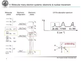
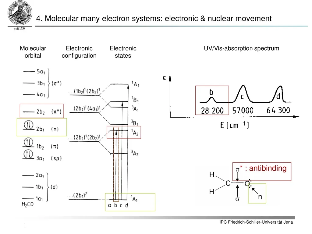
4. Molecular many electron systems: electronic & nuclear movement. Molecular orbital. Electronic configuration. Electronic states. UV/Vis-absorption spectrum. * : antibinding. 4. Molecular many electron systems: electronic & nuclear movement Jablonski-Scheme. v = 1.

E N D
4. Molecular many electron systems: electronic & nuclear movement Molecular orbital Electronic configuration Electronic states UV/Vis-absorption spectrum * : antibinding
4. Molecular many electron systems: electronic & nuclear movement Jablonski-Scheme v = 1 UV-VIS-spectroscopy J = 4 Microwave-spectroscopy S4 Internal conversion[10-14 s] S3 J = 3 Rotational levels Excitation [10-15 s] J = 2 J = 1 Tn J = 0 v = 0 S2 IR- & NIR- spectroscopy Intersystem crossing S1 T1 Vibrational levels v = 4 Fluorescence[10-9 s] Phosphorescence[10-3 s] v = 3 v = 1 v = 0 S0 IPC Friedrich-Schiller-Universität Jena 2
5. UV-Vis-Absorption 5.1 Franck-Condon principle • Interpret electronic absorption spectra based on ||2 of the vibrational levels • electronic transitions (~10-16s) are much faster than the vibrational period (~10-13s) of a given molecule thus nuclear coordinates do not change during transition
5. UV-Vis-Absorption 5.2 Franck-Condon principle • = degree of redistribution of electron density during transition • = degree of similarity of nuclear configuration between vibrational wavefunctions of initial and final states. • Transition probability is proportional to the square modulus of the overlap integral between vibrational wavefunctions of the two electronic states = Franck-Condon-Factor:
5. UV-Vis-Absorption 5.1 Franck-Condon principle |f |i |f |i
5. UV-Vis-Absorption 5.2 Molecular electronic transitions • Molecular electronic transitions: valence electrons are excited from one energy level to a higher energy level. • Electrons residing in the HOMO of a sigma bond can get excited to the LUMO of that bond: σ → σ* transition. • Promotion of an electron from a π-bonding orbital to an antibonding π* orbital: π → π* transition. • Auxochromes with free electron pairs denoted as n have their own transitions, as do aromatic pi bond transitions. • The following molecular electronic transitions exist:σ → σ* π → π* n → σ* n → π* aromatic π → aromatic π* p,p* np* ns* (C=C, C=O) (C=O, C=N, C=S) (–Hal, -S-, -Se- etc.)
5. UV-Vis-Absorption • 5.2 Transition metal complexes • A biologically very important group of metal complex bonds are the porphyrin pigments such as: • Hemoglobin (pigment of the blood, central ion Fe2+) • Cytochromes of respiratory chain • Chlorophyll (green molecules in leaves, central atom Mg) • octahedron structure motive • The four ligand positions of the base of the pyramid are occupied by the lone electron pairs of nitrogen atoms of the plane porphyrin ring system • The two corners of the pyramid are occupied by specific amino acids (histidine) and/or by an oxygen molecule (hemoglobin) Heme-group
5. UV-Vis-Absorption 5.2 Transition metal complexes • Cytochrome C: • Pyramid corners of heme unit are occupied by N-atom of a histidine residue and S-atom of a mezhionine residue • Redox change of cytochromes predominatly occurs at the central iron atom [(Fe2+) ↔ (Fe3+)] a-Peaks= sensitive for redox change(analysis of mitochondria)
° 0,4 A 5. UV-Vis-Absorption • 5.2 Transition metal complexes • Hemoglobin (iron is always found as Fe2+)Arterial oxygen-loaded blood = light redBlood in veines free of oxygen = deep red End-on coordination of O2 (Fe2+ / 75 pm / low spin) B Desoxy Hemoglobin (Fe2+ / 92 pm / high spin)
5. UV-Vis-Absorption 5.3 Polarimetry & Optical rotatory dispersion & Circular dichroism • Fundamental terms: • Polarimetry, optical rotation, circular birefringence: turning of the plane of linearly polarized light • Optically active molecules exhibit different refractive indices for right nR and left nL polarized light nR≠nL • Optical rotatory dispersion (ORD):Wavelength dependency of rotation • Allows determination of absolute configuration of chiral molecules • Circular dichroism:linearly polarized light is transformed into elliptically polarized light upon traveling through matter • Different absorption coefficients for left and right circular polarized light (eR≠eL ).
5. UV-Vis-Absorption 5.3 Polarimetry & Optical rotatory dispersion & Circular dichroism • Polarimetry • What happens if light interacts with chiral molecules? • Enantiomeric molecules interact differently with circular polarized light. • Polarizabilitya depends on direction of rotation of incoming circular polarized light • Optically active substances exhibit different refractive indices for right nRand left nLpolarized light nR ≠ nL
Ey Ex Sample cell Transmitted light Incoming light 5. UV-Vis-Absorption 5.3 Polarimetry & Optical rotatory dispersion & Circular dichroism • Polarimetry: • Linearly polarized light • Different refractive index for its left and right circular constituents • Relative phase shift between left and right • Vector addition yields again linear polarized light with rotated polarization plane phaseshift
5. UV-Vis-Absorption 5.3 Polarimetry & Optical rotatory dispersion & Circular dichroism Polarimetry: Due to the different refractive indices a phase difference d = jL –jR builds up in the active medium which is proportional to the path length l. When exiting the medium linear polarized light where the oscillation plane is rotated by d/2 arises It follows: For a follows: Na-D line l = 589 nm 2-Butanol a = 11.2° (measured value) T = 20°C l = 1dm Difference is rather small! l
5. UV-Vis-Absorption 5.3 Polarimetry & Optical rotatory dispersion & Circular dichroism • Polarimetry • The measured angle-of-rotation results in: • Specific rotation is a substance specific constant (dependent on temperature and wavelength) and is a measure for the optical activity of this particular substance. • Molar rotation is defined as follows: a in angular degree, length in decimeter(!) and c in g ml-1.
5. UV-Vis-Absorption 5.3 Polarimetry & Optical rotatory dispersion & Circular dichroism • Optical rotatory dispersion(ORD) • ORD measures molar rotation [F] as function of the wavelength! • If the substance to be investigated has no electronic absorption within the investigated spectral region the following ORD spectra are obtained • Reason: refractive indices for left and right polarized light change differently with wavelength (rotatory dispersion is proportional to refractive index difference). ORD-spectra of 17ß- and 17a-hydroxy-5a-androstan
5. UV-Vis-Absorption 5.3 Polarimetry & Optical rotatory dispersion & Circular dichroism • Optical rotatory dispersion(ORD) • Refractive indices for left and right polarized light exhibit anomalous dispersion in the range of an absorption band • Cotton effect Positiv negativ Cotton effect
5. UV-Vis-Absorption 5.3 Polarimety & Optical rotatory dispersion & Circular dichroism Optical rotatory dispersion(ORD) ORD-Spektren von 5a-Spirostan und 5a-Spirostan-3-on • Quantitative theoretical correlations between molecular structure and ORD (Cotton effect) are difficult to derive; • Empirical investigation are important: ORD has been successfully applied for constitution elucidation e.g. to position carbonyl groups in complex optically active molecules. • By comparing ORD curves for structurally isomeric ketons (reference material needed!) the keto group can be localized. ORD curve of molecule (2) is a superposition of a negative curve i.e. molecular skeleton without a chromophore (background curve) and a positive Cotton curve (C=O chromophore).
Ey Ex 5. UV-Vis-Absorption 5.3 Polarimetry & Optical rotatory dispersion & Circular dichroism • Circular Dichroism (CD) • Enantiomeric molecules exhibit besides different refractive indices for left and right circular polarized light also different absorption coefficients: • It follows: • left and right circular components ORD : different retardation CD also different absorption • Transmitted light is elliptically polarized. Circular Dichroism
5. UV-Vis-Absorption 5.3 Polarimetry & Optical rotatory dispersion & Circular dichroism Circular Dichroism (CD) • The ratio between short and the long elliptical axis is defined as tangent of an angle , the so called ellipticity (tan = b/a): • a = ER + EL • b = ER - EL • The specific ellipticity is defined as:where 0bs is the experimentally determinedellipticity. • The molar ellipticity is defined as: [10-1 × deg × cm2 × g-1] [10 × deg × cm2 × mol-1]
5. UV-Vis-Absorption 5.3 Polarimety & Optical rotatory dispersion & Circular dichroism Circular Dichroism (CD) • Signal heights are displayed either as absorption difference De or as ellipticity [q]. • Molar ellipticity and circular dichroism can be interconverted: • Correlation between ORD and CD: • ORD is based on the different refractive indices of left and right circular polarized light (nR ≠ nL ) • CD results from the different absorption behavior for left and right circular polarized light (eR ≠ eL) • Connection of both phenomena via Kronig-Kramer relationship: • This relation allows the calculation of an ORD value for a particular wavelength l from the corresponding CD spectrum [q] = [grad cm2 dmol-1]
5. UV-Vis-Absorption 5.3 Polarimetry & Optical rotatory dispersion & Circular dichroism Circular Dichroismus (CD) • Simple model: • For an electronic transition to be CD active the following must be true:µe is the electronic transition dipole moment (= linear displacement of electrons upon transition into an excited state) µm is the magnetic transition moment (= radial displacement of electrons upon excited state transition) • Scalar product is characterized by a helical electron displacement. • Depending on the chirality of the helix preferably more right or left circular polarized light will be absorbed, respectively. Electronic transition Magnetic transition Optical activity
5. UV-Vis-Absorption 5.3 Polarimetry & Optical rotatory dispersion & Circular dichroism Circular Dichroism (CD) • Application field: b-sheet a-helix random coil
5. UV-Vis-Absorption 5.3 Polarimetry & Optical rotatory dispersion & Circular dichroism Circular Dichroism (CD) • Application field: Temperature dependent CD spectra of insuline:For increasing temperature the molecule changes form a-helix into the denaturated random coil form with ß-sheet contributions. Typical reference CD spectra:Poly-L-Lysine in different conformations:a-Helix, b-sheet and random coil.
5. UV-Vis-Absorption 5.3 Polarimetry & Optical rotatory dispersion & Circular dichroism Vibrational-Circular-Dichroism (VCD) • Vibrational transitions in the IR and NIR • VCD monitors difference in absorption between left and right circular polarized light v=1 v=0
5. UV-Vis-Absorption 5.3 Polarimetry & Optical rotatory dispersion & Circular dichroism Vibrational-Circular-Dichroism (VCD) ()-Mirtazapine • Determination of the absoluteconfiguration • Advantages VCD vs. CD • Electronic chromophore is not necessary • VCD exhibits more characterisitic bands
6. Basic concepts in fluorescence spectroscopy v = 1 UV-VIS-spectroscopy J = 4 Microwave-spectroscopy S4 Internal conversion[10-14 s] S3 J = 3 Rotational levels Excitation [10-15 s] J = 2 J = 1 Tn J = 0 v = 0 S2 IR- & NIR- spectroscopy Intersystem crossing S1 T1 Vibrational levels v = 4 Fluorescence[10-9 s] Phosphorescence[10-3 s] v = 3 v = 1 v = 0 S0
6. Basic concepts in fluorescence spectroscopy 6.1 Stokes-Shift = Stokes-Shift due to vibrational energy relaxation within electronic excited state • Energy differences between vibrational states which determine vibronic band intensities are very often the same for ground and electronic excited state • Emission spectrum = mirror image of absorption spectrum • Emission bands are shifted bathochromically i.e. to higher wavelengths