Download
1 / 91
1.43k likes | 2.51k Views
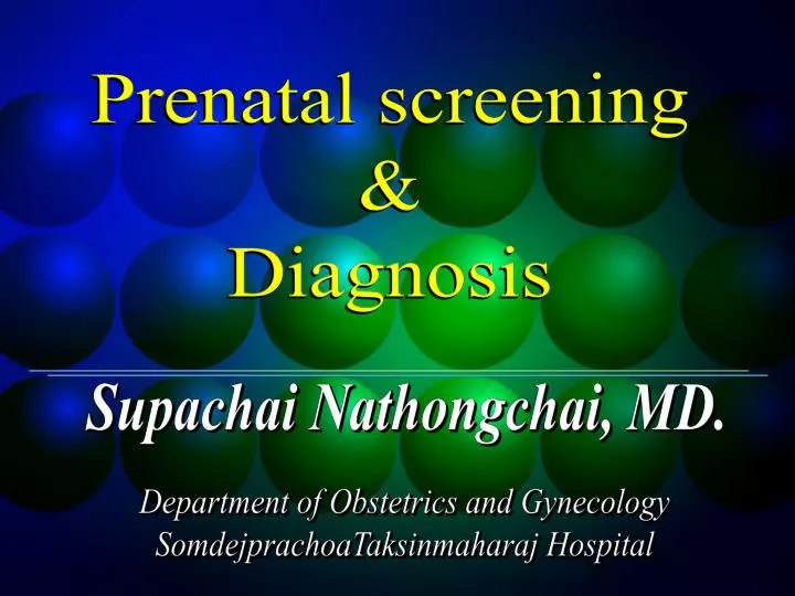
Prenatal screening & Diagnosis. Prenatal screening & Diagnosis. Supachai Nathongchai, MD. Department of Obstetrics and Gynecology SomdejprachoaTaksinmaharaj Hospital. Concept of Screening. Population (pregnant women). Screening test. Screen Positive. Screen Negative.

E N D
Prenatal screening & Diagnosis Prenatal screening & Diagnosis Supachai Nathongchai, MD. Department of Obstetrics and Gynecology SomdejprachoaTaksinmaharaj Hospital
Concept of Screening Population (pregnant women) Screening test Screen Positive Screen Negative • Diagnostic test • Amniocentesis • CVS • Fetal blood sampling No further test
Prenatal Screening • Structural abnormalities • Chromosome abnormalities
Chromosome abnormalities • Trisomy 21 • Trisomy 13 • Trisomy 18 • Monosomy X • Triploidy
Prenatal diagnosis Chorionic villous sampling Amniocentesis Fetal blood sampling Preimplantation genetic diagnosis Chromosome abnormalities But! All methods subject to risk of miscarriage
Screening for Down syndrome • Maternal age • Ultrasonography • Maternal blood tests • Combined
Women < 35 years 70-80% of Down syndrome low risk Neglected
Ultrasonography First trimester screening • Nuchal translucency (NT) • Nasal bone
Observations on an ethnic classification of idiots “ ….. The nose is small. The skin has a slight dirty yellowish tinge, and is different in elasticity, giving the appearance of being too large for the body.” Langdon Down
Nuchal translucency (NT) Measurement of NT
Nuchal translucency (NT) Measurement of NT
Measurement of nuchal translucency The maximum thickness of the subcutaneous translucency between the skin and the soft tissue overlying the cervical spine should be measured by placing the callipers on the lines as shown. During the scan, more than one measurement must be taken and the maximum one should be recorded. + + + + + + + + + +
Measurement of (NT) • CRL 45-84 mm • GA 11-13 wk • Fetus • Sagittal section • Neutral position • Occupies 3/4 of image
Reference range of fetal nuchal translucency with CRL showing the 5th, 25th, 50th, 75th and 95th centiles The 11-14-week scan. The diagnosis of fetal abnormalities
Likelihood ratios for trisomy 21 in relation to the deviation in fetal nuchal translucency thickness The 11-14-week scan. The diagnosis of fetal abnormalities
Recent studies examining the implementation of fetal nuchal translucency screening at 10-14 weeks DR = Detection rate ; FPR = False positive rate
Observations on an ethnic classification of idiots “ ….. The nose is small.The skin has a slight dirty yellowish tinge, and is different in elasticity, giving the appearance of being too large for the body.” Langdon Down
Absence of nasal bone in fetuses with trisomy 21 at 11-14 weeks of gestation Cicero S, et al. The Lancet 2001
Estimated sensitivity and false positive rate for risk cutoffs in screening for trisomy 21 Risk NT screening NT and NB screening cutoffSensitivity (%) FPR (%) Sensitivity (%) FPR (%) 1 in 20 57.36 0.98 81.90 0.62 1 in 35 62.88 1.35 85.89 1.02 1 in 50 65.03 1.76 86.81 1.30 1 in 100 72.09 3.01 88.65 1.64 1 in 150 74.54 4.32 89.26 1.98 1 in 200 77.30 5.71 90.18 2.37 1 in 250 80.67 7.08 91.41 2.68 1 in 300 82.21 8.28 92.02 3.02 1 in 500 85.58 14.00 92.94 4.37 1 in 1000 92.33 27.40 94.7 8.15 Cicero S, et al. The Lancet 2001
Nuchal translucency in twin pregnancies • 448 twin pregnancies • Fetal nuchal translucency thickness was measured by ultrasound examination at 10-14 weeks • Sensitivity and false positive rates of screening for trisomy 21 were calculated • The sensitivity is similar to that in singleton pregnancies • The specificity is lower because translucency is also increased in chromosomally normal monochorionic twin pregnancies Sebire NJ,e t al. Br J Obstet Gynaecol 1996
Fetal karyotyping in twins • Accuracy of obtaining a result from both fetuses • Procedure-related risk of fetal loss • The risks of selective fetocide should the pregnancy be found to be discordant for an abnormality and the parents choose this option In twin pregnancies, selection of the appropriate invasive technique depends on the:
Fetal karyotyping in twins There is an additional advantage of screening by measurement of nuchal translucency thickness; when there is discordancy for a chromosomal abnormality, the presence of a sonographically detectable marker (increased nuchal translucency) helps to ensure the correct identification of the abnormal twin should the parents choose selective termination.
3-D ultrasound measurement of fetal nuchal translucency • It can be used to replicated nuchal translucency measurements only when nuchal skin can also be clearly seen on 2-D ultrasound. Paul C, et al. Ultrasound Obstet Gynecol 2001 • The possibility of rotating a stored volume and inspecting it in three orthogonal planes makes 3-D ultrasound a useful tool for nuchal translucency measurements, especially in doubtful cases. Clementschitsch G, et al. Ultrasound Obstet Gynecol 2001
Conditions associated with increased nuchal translucency Cardiac defects Diaphragmatic hernia Exomphalos Achondrogenesis type II Achondroplasia Asphyxiating thoracic dystrophy Beckwith-Wiedemann syndrome Blomstrand osteochondrodysplasia • Body stalk anomaly • Campomelic dysplasia • EEC syndrome • Fetal akinesia- • deformation sequence • Fryn syndrome • GM1-gangliosidosis • Hydrolethalus syndrome
The second trimester Ultrasound screening of Down syndrome
Genetic Sonogram:Ultrasound indicators for Down 1. Structural anomalies : Cardiac anomaly, DU atresia 2. Soft marker : Echogenic intracardiac foci Pyelectasis, Femur length Humerus Length
Structural anomalies Trisomy 13 Trisomy 18 Trisomy 21 90% 70-80% 20%
Nuchal skin fold thickness Choroid plexus cyst (CPC) Mild ventriculomegaly Nasal bone Echogenic intracardiac foci (EIF) Echogenic bowel Renal pyelectasia Hypoplastic middle phalanx of the fifth digit Soft ultrasound makers
Nuchal skin fold thickness Langdon Down (1866) = thickness of skin of the back Benacerraf et al (1985) = Using as an U/S marker for Down
Cut off threshold of nuchal thickness > 6 mm. 16-20 wks gestation Down abnormal nuchal thickness 40% LR: 17 (CI 8-35) => Expert review is recommended, and karyotyping should be offered Sensitivity 34% Specificity 99.5%
Nuchal thickness : Efficacy Associated with - Congenital heart disease - Genetic syndromes (rare) => Expert review is recommended
Choroid plexus cyst (CPC) Cyst > 3 mm 14 to 24 weeks’ gestation Locate in lateral ventricle Related more to Trisomy 18 Not associated with other fetal abnormalities or abnormal postnatal development.
CPC and Trisomy 18 Snijders et al (1994)Trisomy 18 (58 cases) CPC 50%Normal fetusesCPC 1% Isolated CPCminimal increase risk
Isolated CPC Trisomy 18 LR (trisomy 18) = 7 (CI 4-12) Trisomy 21 Likelyhood ratio = 1.87 (CI 0.78-4.46) Size, Disappearance and Laterality => not increase or reduce risk Gross et al 1995, Gupta et al 1997 , Yoder et al 1999
Recommendation Fetal karyotyping should only be offered if isolated CPCs are found in women 35 years or older or if the maternal serum screen is positive for either trisomy 18 or 21 All women with fetal CPCs and additional malformation should be offered referral and karyotyping All women with CPCs and additional soft markers should be offered additional counselling and further ultrasound review
Mild ventricular dilatation Lateral ventricle : constant along gestation 6.1 + 1.3 mm. Male : female = 6.4 :5.8 mm Definition 10-15 mm. Dangling choroid plexus > 3 mm.from medial wall
Isolate mild ventriculomegaly abnormal fetal karyotype ~ 3.8% (0-28.6%) 0.15% of chromosomally-normal fetuses 1.4% of trisomy 21 fetuses in the second trimester LR 9
Neonatal assessment and follow-up are important to rule out associated abnormalities and are important because of the potential for subsequent abnormal neurodevelopment Cerebral ventricles greater than or equal to 10 mm are associated with chromosomal and central nervous system pathology. Expert review should be initiated to obtain the following: a detailed anatomic evaluation looking for additional malformations or soft markers laboratory investigation for the presence of congenital infection or fetal aneuploidy MRI as a potential additional imaging technique Recommendations
Echogenic intracardiac focus (EIF) A bright focus in papillary muscle Attach to cordae tendinae Move with valvar movement Calcium deposit and surrounded by fibrous tissue Bettelheim et al 1999 Lt ventricle 96% Both ventricle 4.3% Rt ventricle 4.7%
Bromley 1998 EIF : Rt ventricle : Both ventricle Twice the risk Wax + Philput 1998 EIF : Multiple : Dense echo Increase risk
Location of EIF The evidence is best for left or biventricular EICF LR 2.8 (95% CI 1.5-5.5) =>high-risk women. Low-risk => LR 0–1.8 Consensus of the SOGC Imaging and Genetics Committees suggests an LR of 2
EICF should be evaluated as part of the 4-chamber cardiac review during the 16- to 20- week ultrasound Isolated EICF with a fetal aneuploidy risk less than 1/600 by maternal age (31 years) or maternal serum screen requires no further investigations Women with an isolated EICF and a fetal aneuploidy risk greater than 1/600 by maternal age (31 years) or maternal serum screening should be offered counselling regarding fetal karyotyping Women with right-sided, biventricular, multiple, particularly conspicuous, or nonisolated EICF should be offered referral for expert review and possible karyotyping Recommendations
Echogenic bowel echogenicity of abdominal content Compare to liver and bone echo Associated with - Trisomy 21 - Congenital infection - Meconeum ileus (cystic fibrosis) - Bowel atresia - FGR, FDIU
Echogenic bowel Nyberg et al : Grading system Grade 1 : mild echogenic ; diffuse Grade 2 : mod echogenic ; focal Grade 3 : Very echogenic ; bone echo