Download
1 / 110
1.11k likes | 1.18k Views
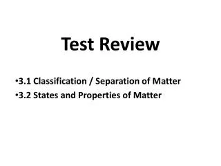
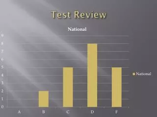
Test Review. Exam 1 Chapters 1 & 4 Spring 2013. Organelle. Atoms. Molecule. Smooth muscle cell. Cellular level Cells are made up of molecules. 2. Chemical level Atoms combine to form molecules. 1. Smooth muscle tissue. Cardiovascular system.

E N D
Test Review Exam 1 Chapters 1 & 4 Spring 2013
Organelle Atoms Molecule Smooth muscle cell Cellular levelCells are made up ofmolecules. 2 Chemical levelAtoms combine to form molecules. 1 Smooth muscle tissue Cardiovascularsystem Tissue levelTissues consist of similartypes of cells. 3 Heart Bloodvessels Blood vessel (organ) Smooth muscle tissue Connective tissue Epithelialtissue Organ levelOrgans are made up of different typesof tissues. 4 Organismal levelThe human organism is made upof many organ systems. Organ system levelOrgan systems consist of differentorgans that work together closely. 6 5 Figure 1.1
Hair Nails Skin (a) Integumentary System Forms the external body covering, and protects deeper tissues from injury. Synthesizes vitamin D, and houses cutaneous (pain, pressure, etc.) receptors and sweat and oil glands. Figure 1.3a
Bones Joint (b) Skeletal System Protects and supports body organs, and provides a framework the muscles use to cause movement. Blood cells are formed within bones. Bones store minerals. Figure 1.3b
Skeletal muscles (c)Muscular System Allows manipulation of the environment, locomotion, and facial expression. Main- tains posture, and produces heat. Figure 1.3c
Brain Nerves Spinal cord (d) Nervous System As the fast-acting control system of the body, it responds to internal and external changes by activating appropriate muscles and glands. Figure 1.3d
Pineal gland Pituitary gland Thyroid gland Thymus Adrenal gland Pancreas Testis Ovary (e) Endocrine System Glands secrete hormones that regulate processes such as growth, reproduction, and nutrient use (metabolism) by body cells. Figure 1.3e
Heart Blood vessels (f) Cardiovascular System Blood vessels transport blood, whichcarries oxygen, carbon dioxide,nutrients, wastes, etc. The heart pumpsblood. Figure 1.3f
Red bone marrow Thymus Lymphatic vessels Thoracic duct Spleen Lymph nodes (g) Lymphatic System/Immunity Picks up fluid leaked from blood vessels and returns it to blood. Disposes of debris in the lymphatic stream. Houses white blood cells (lymphocytes) involved in immunity. The immune response mounts the attack against foreign substances within the body. Figure 1.3g
Nasal cavity Pharynx Bronchus Larynx Trachea Lung (h) Respiratory System Keeps blood constantly supplied with oxygen and removes carbon dioxide. The gaseous exchanges occur through the walls of the air sacs of the lungs. Figure 1.3h
Oral cavity Esophagus Liver Stomach Small intestine Large intestine Rectum Anus (i) Digestive System Breaks down food into absorbable units that enter the blood for distribution to body cells. Indigestible foodstuffs are eliminated as feces. Figure 1.3i
Kidney Ureter Urinary bladder Urethra (j) Urinary System Eliminates nitrogenous wastes from the body. Regulates water, electrolyte and acid-base balance of the blood. Figure 1.3j
Mammary glands (in breasts) Prostate gland Ovary Penis Ductus deferens Testis Uterine tube Scrotum Uterus Vagina (l) Female Reproductive System (k) Male Reproductive System Overall function is production of offspring. Testes produce sperm and male sex hormone, and male ducts and glands aid in delivery of sperm to the female reproductive tract. Ovaries produce eggs and female sex hormones. The remaining female structures serve as sites for fertilization and development of the fetus. Mammary glands of female breasts produce milk to nourish the newborn. Figure 1.3k-l
Necessary Life Functions • Maintaining boundaries between internal and external environments • Plasma membranes • Skin • Movement (contractility) • Of body parts (skeletal muscle) • Of substances (cardiac and smooth muscle)
Necessary Life Functions • Responsiveness: The ability to sense and respond to stimuli (“irritability”) • Withdrawal reflex • Control of breathing rate • Digestion • Breakdown of ingested foodstuffs • Absorption of simple molecules into blood
Necessary Life Functions • Metabolism: All chemical reactions that occur in body cells • Catabolism and anabolism • Excretion: The removal of wastes from metabolism and digestion • Urea, carbon dioxide, feces
Necessary Life Functions • Reproduction • Cellular division for growth or repair • Production of offspring • Growth: Increase in size of a body part or of organism
Survival Needs • Nutrients • Chemicals for energy and cell building • Carbohydrates, fats, proteins, minerals, vitamins • Oxygen • Essential for energy release (ATP production)
Survival Needs • Water • Most abundant chemical in the body • Site of chemical reactions • Normal body temperature • Affects rate of chemical reactions • Appropriate atmospheric pressure • For adequate breathing and gas exchange in the lungs
Homeostasis • Maintenance of a relatively stable internal environment despite continuous outside changes • A dynamic state of equilibrium
Homeostatic Control Mechanisms • Involve continuous monitoring and regulation of many factors (variables) • Nervous and endocrine systems accomplish the communication via nerve impulses and hormones
Components of a Control Mechanism • Receptor (sensor) • Monitors the environment • Responds to stimuli (changes in controlled variables) • Control center • Determines the set point at which the variable is maintained • Receives input from receptor • Determines appropriate response
Components of a Control Mechanism • Effector • Receives output from control center • Provides the means to respond • Response acts to reduce or enhance the stimulus (feedback)
Negative Feedback • The response reduces or shuts off the original stimulus • Examples: • Regulation of body temperature (a nervous mechanism) • Regulation of blood volume by ADH (an endocrine mechanism)
Control Center (thermoregulatory center in brain) Information sent along the afferent pathway to control center Information sent along the efferent pathway to effectors Efferent pathway Afferent pathway Receptors Temperature-sensitive cells in skin and brain Effectors Sweat glands Sweat glands activated Response Evaporation of sweat Body temperature falls; stimulus ends Stimulus Body temperature rises BALANCE Stimulus Body temperature falls Response Body temperature rises; stimulus ends Receptors Temperature-sensitive cells in skin and brain Effectors Skeletal muscles Afferent pathway Efferent pathway Shivering begins Information sent along the efferent pathway to effectors Information sent along the afferent pathway to control center Control Center (thermoregulatory center in brain) Figure 1.5
Negative Feedback: Regulation of Blood Volume by ADH • Receptors sense decreased blood volume • Control center in hypothalamus stimulates pituitary gland to release antidiuretic hormone (ADH) • ADH causes the kidneys (effectors) to return more water to the blood
Positive Feedback • The response enhances or exaggerates the original stimulus • May exhibit a cascade or amplifying effect • Usually controls infrequent events e.g.: • Enhancement of labor contractions by oxytocin (Chapter 28) • Platelet plug formation and blood clotting (Chapter 17)
1 Break or tearoccurs in bloodvessel wall. Positive feedbackcycle is initiated. 3 2 Releasedchemicalsattract moreplatelets. Plateletsadhere to siteand releasechemicals. Positivefeedbackloop Feedback cycle endswhen plug is formed. 4 Platelet plugforms. Figure 1.6
Homeostatic Imbalance • Disturbance of homeostasis • Increases risk of disease • Contributes to changes associated with aging • May allow destructive positive feedback mechanisms to take over (e.g., heart failure)
Anatomical Position • Standard anatomical body position: • Body erect • Feet slightly apart • Palms facing forward
Upper limb Cephalic Acromial Frontal Brachial (arm) Orbital Antecubital Nasal Antebrachial (forearm) Oral Mental Carpal (wrist) Cervical Manus (hand) Thoracic Palmar Axillary Pollex Mammary Digital Sternal Abdominal Lower limb Umbilical Coxal (hip) Pelvic Femoral (thigh) Inguinal (groin) Patellar Crural (leg) Fibular or peroneal Pubic (genital) Pedal (foot) Tarsal (ankle) Thorax Metatarsal Abdomen Digital Back (Dorsum) Hallux (a) Anterior/Ventral Figure 1.7a
Upper limb Cephalic Otic Acromial Occipital (back of head) Brachial (arm) Olecranal Cervical Antebrachial (forearm) Back (dorsal) Manus (hand) Scapular Metacarpal Vertebral Digital Lumbar Lower limb Sacral Femoral (thigh) Gluteal Popliteal Perineal (between anus and external genitalia) Sural (calf) Fibular or peroneal Pedal (foot) Thorax Abdomen Back (Dorsum) Calcaneal Plantar (b) Posterior/Dorsal Figure 1.7b
Body Cavities • Dorsal cavity • Protects nervous system • Two subdivisions: • Cranial cavity • Encases brain • Vertebral cavity • Encases spinal cord
Body Cavities • Ventral cavity • Houses internal organs (viscera) • Two subdivisions (separated by diaphragm): • Thoracic cavity • Abdominopelvic cavity
Cranial cavity Dorsal body cavity Ventral body cavity Cranial cavity (contains brain) Vertebral cavity Superior mediastinum Dorsal body cavity Thoracic cavity (contains heart and lungs) Pleural cavity Pericardial cavity within the mediastinum Vertebral cavity (contains spinal cord) Ventral body cavity (thoracic and abdominopelvic cavities) Diaphragm Abdominal cavity (contains digestive viscera) Abdomino- pelvic cavity Pelvic cavity (contains urinary bladder, reproductive organs, and rectum) (a) Lateral view (b) Anterior view Figure 1.9a-b
Ventral Body Cavities • Thoracic cavity subdivisions: • Two pleural cavities • Each houses a lung • Mediastinum • Contains pericardial cavity • Surrounds thoracic organs • Pericardial cavity • Encloses heart
Serous Membrane (Serosa) • Thin, double-layered membrane separated by serous fluid • Parietal serosa lines internal body walls • Visceral serosa covers the internal organs
Epithelial Membranes • Serous Membranes • Serosae—membranes (mesothelium + areolar tissue) in a closed ventral body cavity • Parietal serosae line internal body walls • Visceral serosae cover internal organs
Parietal peritoneum Parietal pleura Visceral pleura Visceral peritoneum Parietal pericardium Visceral pericardium (c) Serous membranes line body cavitiesclosed to the exterior. Figure 4.11c
Epithelial Membranes • Mucous membranes • Mucosae • Line body cavities open to the exterior (e.g., digestive and respiratory tracts)
Mucosa of nasal cavity Mucosa of mouth Esophagus lining Mucosa of lung bronchi (b) Mucous membranes line body cavitiesopen to the exterior. Figure 4.11b
Diaphragm Liver Right hypochondriac region Left hypochondriac region Epigastric region Stomach Gallbladder Transverse colon of large intestine Ascending colon of large intestine Right lumbar region Left lumbar region Umbilical region Descending colon of large intestine Small intestine Cecum Initial part of sigmoid colon Right iliac (inguinal) region Hypogastric (pubic) region Left iliac (inguinal) region Appendix Urinary bladder (a) Nine regions delineated by four planes (b) Anterior view of the nine regions showing the superficial organs Figure 1.12
Epithelial Membranes • Cutaneous membrane (skin) (More detail with the Integumentary System, Chapter 5)
Tissues • Groups of cells similar in structure and function • Types of tissues • Epithelial tissue • Connective tissue • Muscle tissue • Nerve tissue
Epithelial Tissue (Epithelium) • Two main types (by location): • Covering and lining epithelia • On external and internal surfaces • Glandular epithelia • Secretory tissue in glands
Characteristics of Epithelial Tissue • Cells have polarity—apical (upper, free) and basal (lower, attached) surfaces • Apical surfaces may bear microvilli (e.g., brush border of intestinal lining) or cilia (e.g., lining of trachea) • Noncellular basal lamina of glycoprotein and collagen lies adjacent to basal surface