Download
1 / 17
240 likes | 1.25k Views
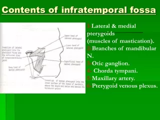
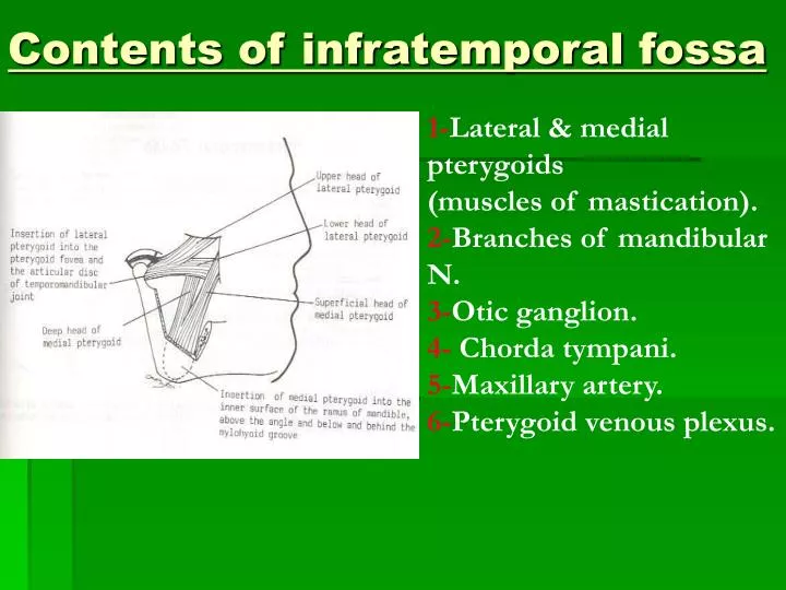
Contents of infratemporal fossa. 1- Lateral & medial pterygoids (muscles of mastication). 2- Branches of mandibular N. 3- Otic ganglion. 4- Chorda tympani. 5- Maxillary artery. 6- Pterygoid venous plexus. Contents of infratemporal fossa : 2-Mandibular Nerve.

E N D
Contents of infratemporal fossa 1-Lateral & medial pterygoids (muscles of mastication). 2-Branches of mandibular N. 3-Otic ganglion. 4- Chorda tympani. 5-Maxillary artery. 6-Pterygoid venous plexus.
Contents of infratemporal fossa : 2-Mandibular Nerve • Origin & course : • It is a mixed N.,formed by 2roots :Large sensory & Small motor roots. • They unite together in foramen ovale to form the main trunk of mandibular N. (mixed). • The main trunk emerge from skull through foramen ovale to reach infratemporal fossa, then divides into a small anterior & a large posterior division.
Branches from main trunk • Meningeal branch (Nervous Spinosus) –sensory nerve :it enters skullthrough foramen spinosum (with middle meningeal artery) to supply meninges in middle cranial fossa. • Nerve to medial pterygoid –motor nerve :It gives off 2 branches, which pass without relay through otic ganglion to supply : tensor tympani (middle ear) & tensor vili palatini (soft palate).
Branches from Anterior Division : • 2 deep temporal nerves (motor) : enter deep surface of temporalis muscle to supply it. • Masseteric nerve (motor). • Nerve to lateral pterygoid muscle (motor). • Buccal nerve ( sensory) : supplies skin over cheek + mucous membrane lining cheek, (it does not supply buccinator muscle, which is supplied by buccal branch offacial nerve).
Branches from Posterior Division :(mainly sensory) 1-Auriculotemporal nerve (sensory) : - It arises by 2 roots that surround middle meningeal artey, then it ascend in company with superficial temporal vessels behind TM joint. - It gives sensory branches to : skin of auricle, external auditory meatus, tympanic membrane, scalp + parotid gland,TM joint. - It gives postganglionic para-sympathetic secretomotor fibres from otic ganglion, to supply parotid gland.
2-Lingual nerve (sensory) :- It arises from posterior division of mandibular nerve, in front of inferioralveolar N. - It lies deep to lateral pterygoid, where it is joined by chorda tympani nerve (branch of facial N. carrying taste ¶sympathetic fibres). - It descends between ramus of mandible & medial pterygoid. -Then, it passes on the inner surface ofthe socket of lower 3rd molar tooth(dangerous area during tooth extraction)- It passes into the submandibular regionsuperficial on the lateralsurface of hyoglossus, here the sub-mandibular ganglion hangs from it.- It ends by dividing into terminal branches to the tongue to carry general & taste sensation from anterior 2/ 3 of m.m of tongue & floor of mouth. Also, it gives secretomotor para-symp.Fs.to submandibular & sublingual glands.
3- Inferior alveolar N. (mixed) :-It is the largest branch of post.division of mandibular N. - It descends on lateral surface ofsphenomandibular ligament. -Then, it enters mandibular canal through mandibular foramen and runs below teeth, supplying the teeth of lower jaw.- Finally it emerges from mental foramen to supply skin of chin (sensory)-N. to mylohyoid (motor) : arises from inferior alveolar N. before it enters the mandibular foramen, it runs in mylohyoid groove of mandible. It ends by supplying mylohyoid m.+ anterior belly of digastric.
3-Otic Ganglion • It is a small parasympathetic ganglion that is functionally associatedwith glosspharyngeal N. • It lies below foramen ovale, medial to mandibular N. • It receives preganglionic para-sympathetic fibres via tympanic branch , tympanic plexus & lesser petrosal N. originate from glossopharyngeal N.(relay in the ganglion). • It sends postganglionic parasympathetic secretomotor fibres via the auriculo-temporal N. to supply the parotid gland.
4-Chorda Tympani • It is a branch of facial N., it leaves the middle ear cavity to enter infratemporal fossa through petrotympanic fissure to join lingual N. • It carries secretomotor parasympathetic fibres to submandibular & sublingual salivary glands. • It carries alsosensory taste fibres continue with lingual N. from anterior 2/3 of tongue & floor of mouth.
5-Maxillary Artery • It arisesbehind to the neck of mandible within the substance of parotid gland, as the larger of the 2 terminal branches ofexternal carotid artery. • It runs upward and forward, superficial to lower head of lateral pterygoid muscle, then it dips between 2 heads of lateral pterygoid to enter pterygopalatine fossa. • Branches :1-inferior alveolar artery : follows inf.alveolar N. into mandibular canal. 2-middle meningeal artery : it passes upward between 2 roots of auriculo-temporal N. it enters skull through foramen spinosum to supply meninges. 3-deep auricular artery : to supply external auditory meatus + tympanic membrane. 4-numerous branches to muscles of mastication.
6-Pterygoid venous plexus • It is a network of veins lying around and inside the substance of lateral pterygoid muscle. • It is drained posteriorly by maxillary v. • It communicates anteriorly with facial vein through deep facial vein. Maxillary Vein • It drains the posterior end of pterygoid venous plexus. • It runs backward with maxillary artery on medial side of neck ofmandible and joins superficial temporal vein within parotid gland to form retromandibular v.
Submandibular Region • It lies under cover of body of mandible, between mandible & hyoid bone. • It contains the following structures : • Muscles :digastric,mylohyoid, hyoglossus, geniohyoid, genioglossus and styloglossus. • Salivary glands : submandibular + sublingual. • Nerves : lingual, glossopharyngeal, & hypoglossal. • Parasympathetic ganglion : submandibular. • Blood vessels : facial & lingual. • Lymph nodes : submandibular.
Muscles of submandibular region : • 1-digastric muscle : • Origin : by 2 bellies -anterior belly : from digastric fossa on the lower border of body of mandible close to symphysis menti. -posterior belly : from medial surface of mastoid process. • Insertion : into the intermediate tendon which is cnnected to hyoid bone by a fibrous loop of deep fascia. This intermediate tendon pierces stylohoid insertion. • Nerve supply : anterior belly : by N.to mylohyoid from mandibular. Posterior belly : by facial N. • Action : depress mandible or elevate hyoid bone during swallowing.
2-mylohyoid muscle : • Origin : flate triangular sheet of muscle arise from mylohyoid line of mandible. • Insertion :the anterior fibres into a median fibrous raphe, the mylohyoid raphe which extends in the median plane from symphysis menti to hyoid bone. The posterior fibres into body of hypoid bone. • Nerve supply : mylohyoid N. from inferior alveolar N. • Action : -they supports tongue & floor of mouth. -they elevate floor of mouth & hyoid bone during 1st stage of swallowing. -they depress mandible (open mouth) when hyoid bone is fixed.
3-hyoglossus muscle : • Origin :upper border of body and greater cornu of hyoid bone. • Insertion : it lies deep to mylohyoid to be inserted to side of posterior ½ of tongue. • Nerve supply : hypoglossal N. • Action :depresses the tongue. 4-styloglossus muscle : • Origin : styloid process. • Insertion : it passes downward on lateral surface of superior constrictor muscle to insert into side of tongue decussating with hyoglossus m. • Nerve supply : hypoglossal N. • Action : retracts the tongue backward.
5-Geniohyoid muscle : • Origin : inferior mental spine, behind symphysis menti of mandible. • Insertion : into anterior surface of body of hyoid bone. • Nerve supply : 1st cervical N. via hypoglossal N. • Action : elevate hyoid bone or depress mandible if hyoid bone is fixed. 6-Genioglossus muscle : it is a fan-shaped m.lies medial to hyoglossus m. • Origin : superior mental spine, behind symphysis menti of mandible. • Insertion : into whole length of tongue + superior fs.into tip of tongue + few inferior fs. Into body of hyoid bone. • Nerve supply : hypoglossal N. • Action : 1-single m. : pulls tongue to opposite side. 2-The 2 ms. Protrude the tongue forward.