Download
1 / 16
230 likes | 619 Views
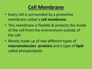
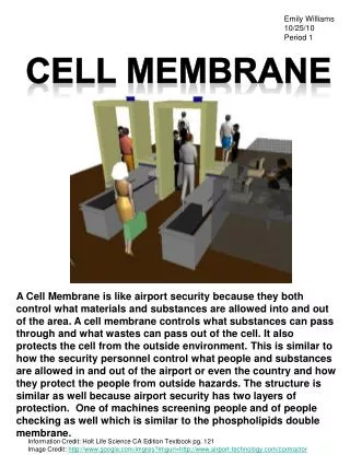
CELL MEMBRANE. Chapter 3 Biology Mr. Gilbertson. CELL MEMBRANE ( PLASMA MEMBRANE). PHOSPHOLIPIDS A GLYCEROL MOLECULE A PHOSPHATE GROUP AND TWO FATTY ACIDS. FAT SANDWICH. CELL MEMBRANE. SEPARATES THE CELL FROM ITS EXTERNAL ENVIRONMENT GIVES SHAPE AND FLEXIBILITY TO CELL

E N D
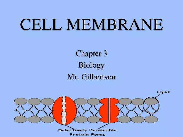
CELL MEMBRANE Chapter 3 Biology Mr. Gilbertson
PHOSPHOLIPIDS A GLYCEROL MOLECULE A PHOSPHATE GROUP AND TWO FATTY ACIDS
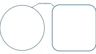
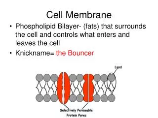
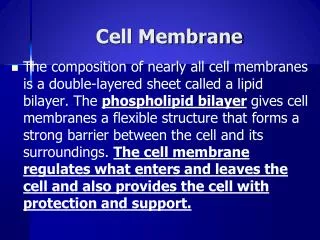
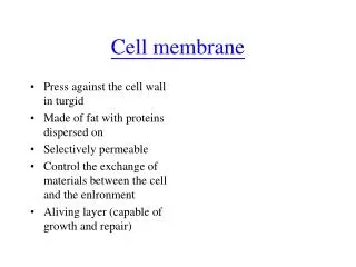
CELL MEMBRANE • SEPARATES THE CELL FROM ITS EXTERNAL ENVIRONMENT • GIVES SHAPE AND FLEXIBILITY TO CELL • COMPLEX BARRIER ALLOWING SOME MOLECULES IN YET NOT ALLOWING OTHER TO PASS • SELECTIVELY PERMEABLE MEMBRANE • 7.5 TO 10 NANOMETERS THICK
Selectively Permeable Membrane Allows some but not all molecules to pass Permeability changes with needs of the cell
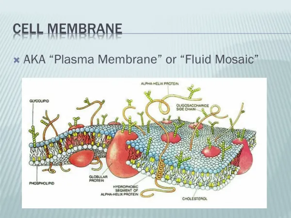
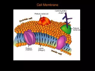
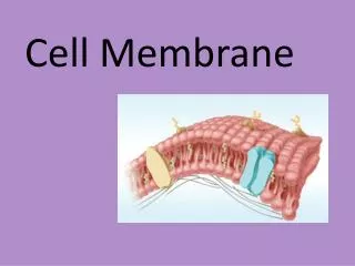
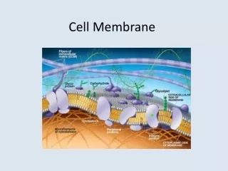
FLUID MOSAIC MODELPLASMA MEMBRANE BILIPID LAYER WITH GLOBULAR GLYCOPROTEINS FLOATING WITHIN A SEA OF LIPIDS
FLUID MOSAIC MODEL • MEMBRANE IS COMPOSED OF TWO LAYERS OF LIPIDS • HYDROPLILLIC END TO THE OUTSIDE OF MEMBRANE • HYDROPHOBIC END TO THE INSIDE OF THE MEMBRANE • PROTEINS ARE EMBEDDED WITHIN THE LIPID LAYERS • SOME ARE ASSOCIATED WITH THE INNER MEMBRANE SOME ARE ASSOCIATED WITH THE OUTER MEMBRANE • SOME PENETRATE THE ENTIRE MEMBRANE • LIPIDS ARE FLUID AND CAN MOVE ABOUT FREELY • PROTEINS ARE ALSO FREE TO MOVE ABOUT SO THE MOSAIC OR “PATTERN” CHANGES
Fluid Mosaic Membrane ModelA sea of lipids with associated glycoproteins constantly moving an changing positions or pattern
RECEPTORS RECEPTORS ARE LOCATED IN THE MEMBRANE, THEY DETECT SIGNAL MOLECULES CALLED LIGANDS IN THE CELLS ENVIRONMENT THAT INDICATE A CHANGE AND PERFORM AN APPROPRIATE RESPONSE.
Types of Receptors Intracellular receptors • Molecules cross the membrane and bind to receptor inside the cell • Most of these molecules are nonpolar allowing passage through the membrane (hormones) • These molecules only produce an effect on a particular cell if the appropriate receptor is present within the cell, in others cells they have no effect.
Receptor binds to ligand and both can now pass easily through the membrane.Once inside the ligand (hormone) triggers a reaction which cause a protein to be produced
Types of Receptors Membrane Receptors • Molecules that cannot cross the membrane may bind to a receptor in the membrane. • The presence of the ligand causes the receptor to change shape, allowing specific molecules into the cell or causing molecules in the cell to respond. • This sets in motion a complex series of events within the cell (elimination of wastes)
Membrane Receptorsbind to a ligand and cause a cascade of events leading to some change within the cell.
AN EXAMPLE OF A RECEPTOR IN ACTION IF INSULIN IS PRESENT IT (A HORMONE LIGAND) BONDS WITH A RECEPTOR, THIS CAUSES THE RECEPTOR TO OPEN A CHANNEL THROUGH WHICH GLUCOSE CAN ENTER THE CELL. IF INSULIN IS NOT PRESENT THE CHANNEL REMAINS CLOSED AND NO GLUCOSE CAN ENTER THE CELL. HIGH GLUCOSE IN THE SUGAR TRIGGERS PRODUCTION OF INSULIN IN NON-DIABETICS.