Download
1 / 36
430 likes | 804 Views
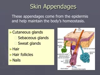
Skin and Its Appendages. Chapter 6. Integument. Skin is the largest organ in the body. Makes up 16% of total body weight. Approximately 1.6-1.9 m2 in the average sized adult Integumentary system describes the skin and its appendages Hair Nails Skin glands. Structure of Skin.

E N D
Skin and Its Appendages Chapter 6
Integument • Skin is the largest organ in the body. Makes up 16% of total body weight. • Approximately 1.6-1.9 m2 in the average sized adult • Integumentary system describes the skin and its appendages • Hair • Nails • Skin glands
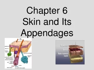
Structure of Skin • Classified as a cutaneous membrane • Two primary layers- dermis and epidermis joined by a dermal epidermal junction • Subcutaneous layer- hypodermis or superficial fascia lies beneath the dermis • Thick and thin skin • Thick skin- soles and palms (4-5mm thick) • Thin skin- covers most of the body (1-3mm thick)
Epidermis- Cell types • Keratinocytes- make up 90% of cells present- principal structural element of outer skin • Melanocytes- pigment producting cells (%5 of total) give color and filter UV light • Langerhans cells- play a role in immune response
Cell Layers of Epidermis 1. Stratum Corneum (top layer) • Dead cells filled with keratin (barrier area) 2. Stratum Lucidum (clear layer) cells filled with keratin precursor called eleidin absent in thin skin. 3. Stratum granulosum (granular layer) cells arranged 2-3 layers and filled with keratohyalin granules that contain a high # of lysosomes ( to digest the cytoplasm as it is replaced with keratin)
Cell layers- epidermis continued 4. Stratum Spinosum (spiny layer) cells arranged in 8-10 layers with prominent desmosomes (strong connections between cells appear spiny in microscope): Rich in RNA which is necessary for the protein synthesis of Keratin. 5. Stratum basale (base layer) single layer of columnar cells: only these cells undergo mitosis and then migrate through the other layers until they are shed.
Epidermal growth and repair • Turnover or regeneration time refers to time it takes for cells to go from base layer to corneum. This takes about 35 days • Shortened turnover time can be created with abrasion. However prolonged abrasion of an area that stimulates mitotic division results in a callus (increased stratum corneum) • Normally 10-12% enter mitosis daily in the stratum basale.
Dermis Epidermis junction • A definite basement membrane, specialized fibrous elements and a polysaccharide gel serve to “glue” the epidermis to the dermis. • The junction serves as a partial barrier to the passage of some cells and large molecules
Dermis “TRUE SKIN” • Much thicker • Gives strength to the skin • Reservoir storage area for water and electrolytes • Contains specialized sensory receptors, muscle fibers hair follicles, sweat and sebaceous glands and blood vessels.
Dermis cont… • Rich vascular supply plays a critical role in the regulation of body temperature. • As the body senses an increase in temperature the blood vessels of the skin dilate and heat radiates out of the blood vessels through the dermis (vasodilation) • As the body senses a decrease in temperature the blood vessels will constrict (vasoconstriction). This keeps warmer blood circulating deeper in the tissue in more “critical” areas.
Layers of the Dermis • Papillary layers- composed of dermal papillae that project into the epidermis; contains fine collagenous and elastic fibers; contains the dermal epidermal junction ; forms a unique pattern that gives individual fingerprints
Layers of dermis cont… • Reticular layer- contain dense, interlacing white collagenous fibers and elastic fibers to make the skin tough yet stretchable; when processed from animal skin it produces leather.
Dermal Growth and Repair • The dermis does not continually shed and regenerate itself as does the epidermis • During wound healing the fibroblasts begin forming and unusually dense mass of new connective fibers; if not replaced by normal tissue, this mass remains a scar. • Langer’s cleavage lines patterns formed by the collagenous fibers of the reticular layer of the dermis. Important for surgeons • If the elastic fibers of the dermis are stretched too much as in obesity or pregnancy or rapid growth phases the result is tiny linear markings or stretch marks- actual tears in the elastic
Skin Color • Basic determinant is quantity of melanin, found in the stratum basale of the epidermis in all races. • Melanin is formed from melanocytes from the amino acid tyrosine. • Albinism- congenital absence of melanin.
Skin color cont… • Process of skin coloration is regulate by tyrosinase(enzyme that breaks down tyrosine) , exposure to sunlight, and certain hormones including ACTH (adrenocorticotropic hormone) that regulates aging. • Carotene (yellowish color) can also contribute to skin color • Color changes also occur as a result of changes in blood flow to skin and circulating levels of unoxygenated hemoglobin. (bluish to pale appearance)
Functions of the skin • Protection • Prevent dehydration • UV barrier through melanin • Surface film- sweat, oil, epithelial cells that are shedding • Antibacterial, antifungal activity • Lubrication • Hydration of skin surface • Buffer caustic agents • Blockade of toxic agents
Functions of skin cont. • Sensation- sophisticated sense organ • Able to detect temperature, pressure, touch , pain and other general senses. http://www.innerbody.com/image_nerv16/nerv141.html • Movement without injury due to elastic nature of skin • Excretion of water, urea, ammonia, and uric acid • Vitamin D production • Exposure to UV light converts 7 dehydorCHOLESTEROL to cholecalciferol (precusor to vit D) • Vitamin D is classified as a hormone once process is complete
Functions of skin • Immunity • Phagocytic cells destroy bacteria • Langerhans cells trigger helpful immune reaction with helper T cells. Homeostasis of body temperature • Heat production must equal heat lost for temperature to be balanced • Heat production- metabolism of foods in skeletal Muscle and liver. Chief determinant is the amount of muscular work being done.
Function of skin: homeostasis of body temp • 3. Heat Loss- approximately 80% of heat loss occurs through the skin; remaining 20% occurs through the skin mucosa of the respiratory, digestive, and urinary tracts. • Evaporation- to evaporate fluid from skin energy must be expended (sweat) • Radiation- heat loss in cool environmental conditions • Conduction-very small amounts of heat loss • Convection- transfer of heat away from skin by movement of air (sitting under a fan or air conditioner) • Fig 6-6 pg 173
Homeostatic regulation of heat loss • Heat loss is controlled by a negative feed back loop • Hypothalamus senses temperature change from its “set point” • If too high- signal sent to sweat glands and blood vessels • If too low- to muscles to shiver to create frictional heating • Once temperature is returned to set point nervous response is inhibited.
BURNS- figure 6-9 p 176 • Injury or death to skin cells caused by heat, UV, electrical current or corrosive chemical • Severity is determined by depth of lesion and % of body surface burned • Estimating burn area • RULE OF PALMS- palm is approximately 1% of body surface • Lund-Browder Charts- more accurate with children due to the difference in proportions of head to body.
Burns- Rule of NINES • Head/neck - 9% • Each arm - 9% • Anterior thorax - 18% • Posterior thorax - 18% • Each leg - 18% • Perineum - 1%
First degree burn • Minor pain – no significant tissue destruction • Some reddening of the skin • No blistering, bust some peeling of surface occurs • No scarring • Typical sunburn • Partial thickness burn
Second degree burn • Severe pain • Damage or destruction of epidermis and upper dermal layers • Blisters form with swelling and edema (fluid in the tissue) • Dermal tissue death not complete but scarring common • Partial thickness burn
Third degree burn- full thickness burn • Total destruction of both epidermis and upper dermal layers • Tissue death extends below level of hair follicles and sweat glands • No immediate pain; nerve endings are destroyed • Burning may involve deep tissues , including muscle and bone • Scarring is a serious problem
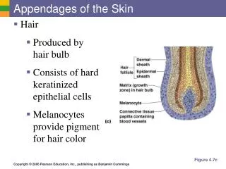
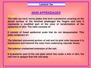
Appendages of the Skin • Hair • Papilla- capillaries clustered under follicle • Root- tip of hair embedded in follicle • Shaft- visible part of hair • Medulla- inner core of hair: Cortex- outer portion • Color- results from the different amounts of melanin in cortex of hair • Growth- periods of growth and rest. Grows about 5 inches per year. • Sebaceous glands- oil glands that secrete sebum into follicle • Male pattern baldness
Appendages cont… • Nails • Epidermal cells converted to hard keratin • Nail body – visible part of each nail • Root- part of nail in groove hidden by fold of skin, cuticle • Lunula- moon-shaped white area of nearest root • Nail bed- layer of epithelium under nail body- abundant blood vessels • Growth average: 0.5mm per week or 1 inch per year
Appendages cont. . . SKIN GLANDS • Two types of sweat glands • Eccrine glands • Most numerous sweat glands, quite small • Over total body surface • Simple coiled tubular glands • Function entire life • Help body maintain temperature • http://www.innerbody.com/image_nerv16/nerv141.html
Sweat Glands cont… • Apocrine glands • Located deep in subcutaneous layer • Limited distribution- axilla, areola of breast, anus • Large 5mm in diameter • Simple branched tubular glands • Function at puberty • Secretion cyclic changes in female with menstrual cycle
Skin Glands- Sebaceous glands • Secrete sebum- oily substance that keeps hair and skin soft and pliant; prevents water loss • Lipid components have antifungal activity • Simple branched glands • In dermis except for palms and soles • Secretion increases in adolescence forming pimples, blackheads
Ceruminous glands • Modified apocrine sweat glands • Simple coiled, tubular glands • Empty contents into external ear canal alone with sebaceous glands • Mixed secretions result in ear wax • Functions to protect area from dehydration. • Excess production can block ear canal and cause loss of hearing
Cycle of Life: skin • Children • Smooth skin, elasticity, and flexibility • Few sweat glands • Rapid healing • Adult • Activation of sweat and sebaceous glands • Increased sweat and body odor • Increased sebum production (oil)= acne • Old age • Decreased sweat and sebaceous activity • Wrinkling • Decreased in the body’s ability to cool itself