Download
1 / 21
210 likes | 370 Views
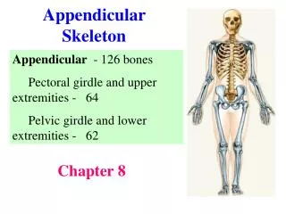
Appendicular Skeleton. Lower Extermities. Pelvic Girdle. Identify the bones of the pelvic girdle and their principal markings. This consists of the two hip bones also called coxal bones. They are united to each other anteriorly at a joint called the pubic symphysis.

E N D
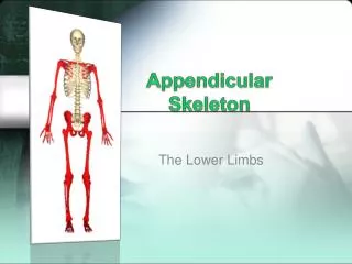
Appendicular Skeleton Lower Extermities
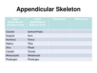
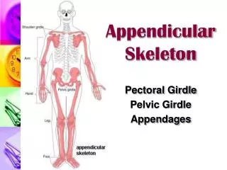
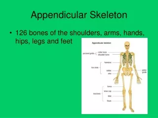
Pelvic Girdle • Identify the bones of the pelvic girdle and their principal markings. • This consists of the two hip bones also called coxal bones. • They are united to each other anteriorly at a joint called the pubic symphysis. • They unite posteriorly with the sacrum at the sacroiliac joint. • This complete ring-bony pelvis
Pelvic Girdle • Each of the two hip bones of a newborn consists of three bones separated by a cartilage-a superior ilium, an inferior and anterior pubis and an inferior and posterior ischium. • Eventually the three separate bones fuse together.
Ilium • This is the largest of the three components. • Divided into superior ala an inferior body, enters into formation of acetabulum. • Superior border or iliac crest ends in a blunt anterior superior iliac spine. • Below the spine is the anterior inferior iliac spine.
Ilium • Below the posterior inferior iliac spine is the greater sciatic notch, through which the sciatic nerve passes. • The medial surface of the ilium contains the iliac fossa. • Posterior to this fossa are the iliac tuberosity • auricular surface that articulates with the sacrum to form sacroiliac joint.
Ilium • A ridge called the arcuate line. • Three arched lines-posterior gluteal line, the anterior gluteal line and the inferior gluteal line. • The tendons of the gluteal muscles attach to the ilium between these lines.
Ischium • This is the inferior, posterior portion iof the hip bone. • It is composed of a superior body and inferior ramus which joins the pubis. • It contains the ischial spine, a lesser sciatic notch, and a rough ischial tuberosity. • Together the ramus and pubis surround the obturator foramen. (largest foramen)
Pubis • The pubis or os pubis is the anterior and inferior part of the hip bone. • It consists of a superior ramus, an inferior ramus and a body between the rami. • Anterior border of the body-pubic crest and alateral projection called pubic tubercle. • The iliopectineal line.
Pubic Symphysis • This is the joint between the the two hip bones. • The acetabulum is the deep fossa that is formed by the ilium, ischium and pubis. • Together the acetabulum and the femoral head form the hip (coxal) joint. • Acetabulaur notch-forms a foramen.
True and False Pelves • Divided into superior and inferior portions by a boundary called-pelvic brim
Comparison of Male and Female Pelves • Bones of a male are larger and heavier. • Larger surface markings • female’s pelvis is wider and shallower than the male.
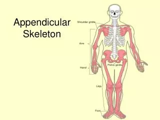
Lower limb • Identify the bones and their principal markings. • The two lower limbs are each composed of 30 bones. • Each lower limb includes the femur (thigh), the patella (kneecap), tibia and fibula, tarsals of the tarsus (ankles), metatarsals and phalanges.
Femur • Strongest, heaviest and longest bone. • Its proximal end articulates with the acetabulum. • Distal end articulates with the tibia and kneecap. • The body of the femur angles medially. • Greater angle of convergence in the females.
Femur • The proximal end consists of head that articulates with acetabulum to form hip (coxal) joint. • Greater trochanter and lesser trochanter. • Intertrochanteric line • gluteal tuberosity and linea aspera • medial epicondyle and lateral epicondyle • intercondylar fossa
Patella • A small triangular bone located anterior to the knee joint. • The broad superior end is called the base and the pointed inferior end is called apex. • Articular facets • patellofemoral joint intermediate component of the tibiofemoral joint • increases leverage, protects, maintains position.
Tibia • The tibia or shin bone is the larger, medial weight bearing bone. Articulates at its proximal end with the femur and fibula and distal with the fibula and talus bone of the ankle.
Tibia • Markings • lateral condyle • medial condyle • tibiofemoral joint • intercondylar eminence • tibial tuberosity • medial malleolus • fibular notch-distal tibiofibular joint
Fibula • Parallel and lateral to tibia, but considerably smaller. • Markings • head • proximal tibiofibular joint • lateral malleolus
Tarsals • Proximal region of the foot and conssists of 7 tarsal bones. • Talus • calcaneus • cuboid • navicular • 3 cuneiform bones • intertarsal joints • talocrural joint
Metatarsals • Intermediate region of the foot. • Consists of five metatarsal bones-I to V • consists of a proximal base, a medila shaft and a distal head. • Tarsometatarsal joint • distally-metatarsophalangeal joints
Phalanges • Resemble those of the hand. • Numbered I to V from the big toe. • Proximal base, an intermediate shaft and a distal head. • The great big toe or hallux. Has two phalanges. • All the rest have three. • Interphalangeal joints.