Download
1 / 1
10 likes | 147 Views
PEG hydrogels functionalized with a Collagen-mimetic peptide and vascular endothelial growth factor for regeneration of critically-sized bone defects. 4-arm PEG-MAL. Functionalized Precursor. Cross-linked Network. Small Degradation Products. Cross-linker. pH 6.5-7.5. Ligand. HS--. --SH.

E N D
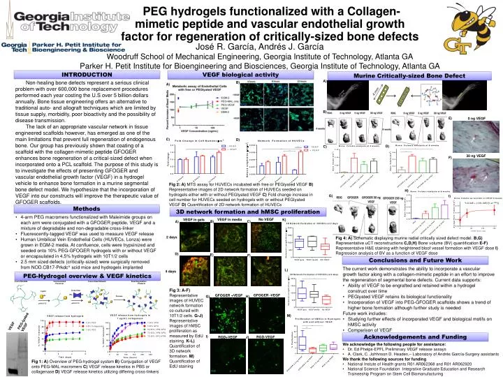
PEG hydrogels functionalized with a Collagen-mimetic peptide and vascular endothelial growth factor for regeneration of critically-sized bone defects 4-arm PEG-MAL Functionalized Precursor Cross-linked Network Small Degradation Products Cross-linker pH 6.5-7.5 Ligand HS-- --SH --SH N N N N pH 6.5-7.5 José R. García, Andrés J. García Woodruff School of Mechanical Engineering, Georgia Institute of Technology, Atlanta GA Parker H. Petit Institute for Bioengineering and Biosciences, Georgia Institute of Technology, Atlanta GA Protease VEGF biological activity INTRODUCTION Murine Critically-sized Bone Defect A) Non-healing bone defects represent a serious clinical problem with over 600,000 bone replacement procedures performed each year costing the U.S over 5 billion dollars annually. Bone tissue engineering offers an alternative to traditional auto- and allograft techniques which are limited by tissue supply, morbidity, poor bioactivity and the possibility of disease transmission. The lack of an appropriate vascular network in tissue engineered scaffolds however, has emerged as one of the main limitations that prevent full regeneration of endogenous bone. Our group has previously shown that coating of a scaffold with the collagen-mimetic peptide GFOGER enhances bone regeneration of a critical-sized defect when incorporated onto a PCL scaffold. The purpose of this study is to investigate the effects of presenting GFOGER and vascular endothelial growth factor (VEGF) in a hydrogel vehicle to enhance bone formation in a murine segmental bone defect model. We hypothesize that the incorporation of VEGF into our constructs will improve the therapeutic value of GFOGER scaffolds. B) A) B) E) 0 ng VEGF C) D) D) C) 30 ng VEGF F) Fig 2: A) MTS assay for HUVECs incubated with free or PEGlyated VEGF B) Representative images of 2D network formation of HUVECs seeded on hydrogels either with or without PEGlyated VEGF C) Fold change increase in cell number for HUVECs seeded on hydrogels with or without PEGlyated VEGF D) Quantification of 2D network formation of HUVECs H) G) I) Y=0.045 + (.119-.045)*(1-e(-.039x)) Methods 3D network formation and hMSC proliferation • 4-arm PEG macromers functionalized with Maleimide groups on each arm were conjugated with a GFOGER peptide, VEGF and a mixture of degradable and non-degradable cross-linker • Fluorescently-tagged VEGF was used to measure VEGF release • Human Umbilical Vein Endothelial Cells (HUVECs, Lonza) were grown in EGM-2 media. At confluence, cells were trypsinized and seeded onto 10% PEG-GFOGER hydrogels with or without VEGF or encapsulated in 4.5% hydrogels with 10T1/2 cells • 2.5 mm sized defects (critically-sized) were surgically removed from NOD.CB17-Prkdc^ scid mice and hydrogels implanted No VEGF VEGF in media VEGF in gels K) B) C) A) 2 days Fig 4: A) Schematic displaying murine radial critically sized defect model. B,G) Representative uCT reconstructions C,D,H) Bone volume (BV) quantification E-F) RepresentativeH&E staining with heightened bloof vessel formation with VEGF dose I) Regression analysis of BV as a function of VEGF dose CD31 CD31 CD31 SMA SMA SMA D) E) F) Conclusions and Future Work • The current work demonstrates the ability to incorporate a vascular growth factor along with a collagen-mimetic peptide in an effort to improve the regeneration of segmental bone defects. Current data supports: • Ability of VEGF to be engrafted and retained within a hydrogel construct over time • PEGlyated VEGF retains its biological functionality • Incorporation of VEGF into PEG-GFOGER scaffolds shows a trend of higher bone formation although further study is needed • Future work includes: • Studying further effects of incorporated VEGF and biological motifs on hMSC activity • Comparison of VEGF L) 4 days PEG-Hydrogel overview & VEGF kinetics CD31 CD31 CD31 SMA SMA SMA Fig 3:A-F) Representative images of HUVEC network formation co-cultured with 10T1/2 cells. G-J) Representative images of hMSC proliferation as measured by EdU staining. K-L) Quantification of 3D network formation. M) Quantification of EdU staining GFOGER -VEGF GFOGER +VEGF G) H) M) PEG-lyated VEGF VEGF EdU EdU Acknowledgements and Funding I) RGD-VEGF J) RGD+VEGF • We acknowledge the following people for assistance: • Dr. Ed Phelps-EPFL Preliminary VEGF release assays • A. Clark, C. JohhnsonD. Headen,– Laboratory of Andrés García-Surgery assistants • We thank the following sources for funding • National Instute of Health grants R01 AR062368 and R01 AR062920 • National Science Foundation Integrative Graduate Education and Research Traineeship Program on Stem Cell Biomanufacturing Fig 1: A) Overview of PEG-hydrogel system B) Conjugation of VEGF onto PEG-MAL macromersC) VEGF release kinetics in PBS or collagenase D) VEGF release kinetics utilizing differing cross-linkers EdU EdU