Download

1 / 27
410 likes | 904 Views
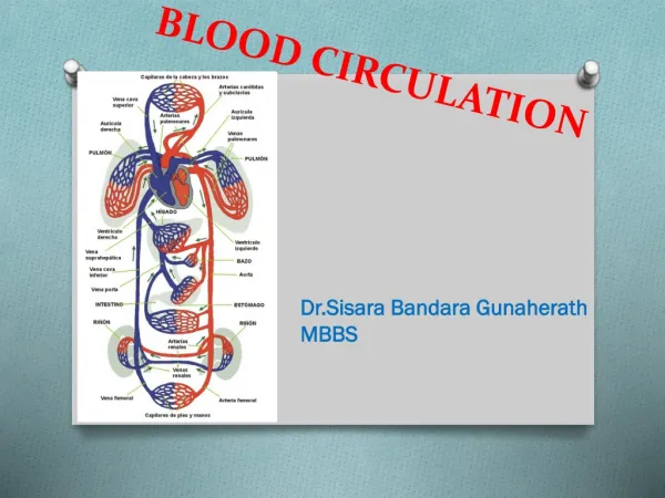
Cerebral Blood Circulation. OBJECTIVES. At the end of the lecture, students should be able to: List the cerebral arteries. Describe the cerebral arterial supply regarding the origin, distribution and branches. Describe the arterial Circle of Willis .

E N D
OBJECTIVES At the end of the lecture, students should be able to: • List the cerebral arteries. • Describe the cerebral arterial supply regarding the origin, distribution and branches. • Describe the arterial Circle of Willis . • Describe the cerebral venous drainage and its termination. • Describe arterial & venous vascular disorders and their clinical manifestations.
Large mass of nervous tissue located in the cranial cavity. Has four major regions. Review: THE BRAIN Cerebrum (Cerebral hemispheres) Diencephalon: Thalamus, Hypothalamus, Subthalamus & Epithalamus Cerebellum Brainstem: Midbrain, Pons & Medulla oblongata
Review: CEREBRUM • The largest part of the brain, and has two hemispheres. • The surface shows elevations called gyri, separated by depressions called sulci. • Each hemispheresdivided into 4 lobes by deeper grooves. • Lobs are separated by deep grooves called fissures.
CEREBRAL ARTERIAL SUPPLY • It is Provided by Two Systems: • Carotid System • Vertebro Basilar System
CEREBRAL ARTERIAL SUPPLY • The two Systems join at Circle of Willis, • It is Located on the base of the brain • It Encircles: • Optic chiasma • Hypothalamus • Midbrain
CIRCULUS ARTERIOSUS (OF WILLIS) Named after Thomas Willis (1621–1675), an English physician • It is Formed by: • Two Anterior cerebral arteries • Two Internal carotid arteries • Two Posterior cerebral arteries • Two Posterior communicating arteries • One Anterior communicating artery • It Gives numerous small vessels that penetrate the surface of the brain • Perforating arteries • They are divided into: • Anterior perforating arteries • Posterior perforating arteries
ANTERIOR PERFORATING ARTERIES • Arise from: • Anterior cerebral artery, • Anterior communicating artery • Middle cerebral artery • Enter brain through: • Anterior perforated substance • irregularly quadrilateral area in front of the optic tract and behind the olfactory trigone. • Supply: • Large part of basal ganglia • Optic chiasma • Internal capsule • Hypothalamus
POSTERIOR PERFORATING ARTERIES • Arise from: • Posterior cerebral artery • Posterior communicating artery • Enter brain through: • Posterior Perforated substance • Supply: • Ventral Portion of Midbrain • Parts of Subthalamus and Hypothalamus
ANTERIOR CEREBRAL ARTERY • Supplies: Orbital and medial surfaces of frontal and parietal lobes
MIDDLE CEREBRAL ARTERY • Supplies: Entire Superolateral surface: • Somatosensory Cortex • Motor Cortex • Broca's Area • linked to speech production. • Heschl’s Gyrus • to process incoming auditory information • Wernicke’s Area • It is involved in the understanding of written and spoken language
POSTERIOR CEREBRAL ARTERY • Supplies: • Anterior and inferior temporal lobes • Uncus • involved in olfactory processing and emotional, associational processing • Inferior temporal gyri • Inferior and Medial Occipital lobe
DISTRIBUTION OF CEREBRAL ARTERIES MCA PCA ACA
ARTERIAL DISORDER • Stroke • Sudden occlusion • Hemorrhage • Aneurysm • is a localized, blood-filled balloon-like bulge in the wall of a blood vessel. • Angioma • benign tumors derived from cells of the vascular or lymphatic vessel walls (epithelium) or derived from cells of the tissues surrounding these vessels.
ACCLUSION OF ACA • Manifestations: • Motor disorder in contralateral distal leg • Difficulty in Prefrontal lobe Functions: • Cognitive thinking • Judgment • Motor initiation • Self monitoring
ACCLUSION OF MCA • Manifestations: • Contralateral weakness of: • face, arm, and hand more than legs • Contralateral sensory loss of: • face, arm, and hand more than legs • visual field cut (damage to optic radiation) • Aphasia: language disorders • Broca's: production • Wernicke's: comprehension
ACCLUSION OF PCA • Manifestations: • Visual disorder • Contralateral homonymous hemianopsia • Bilateral lesions: cortical blindness • Memory impairment • If temporal lobe is affected
CEREBRAL VENOUS DRAINAGE • It involves: • Superficial (cortical) veins: • Drain the cortical surface • Deep veins: • Drain the deep structures • These veins ultimately drain into: • Dural Venous Sinuses • The Veins are thin walled and are devoid of valves.
SUPERFICIAL CORTICAL VEINS • Lie on the brain surface, in the Subarchnoid space. • They are divided into: • Superior cerebral veins • Inferior cerebral veins • Superficial middle cerebral vein
SUPERFICIAL CORTICAL VEINS • Superior Cerebral Veins • 6 to 12 veins • Drain lateral surface of brain above the lateral sulcus • Terminate mainly into the Superior Sagittal sinus, and partly into superficial middle cerebral vein.
SUPERFICIAL CORTICAL VEINS • Inferior Cerebral Veins • Run below the lateral sulcus • Drain the lateral surface of the temporal lobe • Terminate partly into superficial middle cerebral vein & partly into Transverse sinus.
SUPERFICIAL CORTICAL VEINS • Superficial Middle Cerebral Vein • Runs along the lateral sulcus • Terminates into the Cavernous sinus • Connected posteriorly by Superior & Inferior anastomotic veins to Superior Sagittal & Transverse sinuses respectively.
DEEP CEREBRAL VEINS • They drain the internal structures; • Basal ganglia • Internal capsule • Thalamus • They merge to form the Internal Cerebral Veins. • The two veins unite in the midline to form the Great Cerebral vein. • This short vessel is continuous with the Straight Sinus.
DURAL VENOUS SINUSES Paired Single • Transverse • Sigmoid • Cavernous • Petrosal • Superior sagittal • Inferior sagittal • Straight • Occipital Blood flows from transverse & sigmoid sinuses into IJV
VENOUS DISORDER • Infarction • refers to tissue death (necrosis) that is caused by a local lack of oxygen due to obstruction of the tissue's blood supply • Sinus thrombosis: • SSS thrombosis • Superior Sagittal Sinus • Can complicates ear infection • Cavernous Sinus thrombosis • As a complication of infection in the dangerous area of the face • Obstruction of venous drainage of the brain leads to Cerebral swelling (edema) and raised Intracranial Pressure.