Download
1 / 108
1.56k likes | 3.53k Views
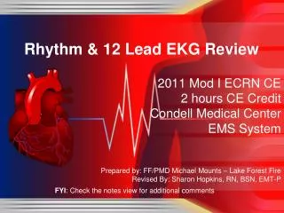
Rhythm & 12 Lead EKG Review. March 2011 CE Condell Medical Center EMS System Site code # 107200E-1211 Prepared by: FF/PMD Michael Mounts – Lake Forest Fire Revised By: Sharon Hopkins, RN, BSN, EMT-P. Objectives. Upon successful completion of this module, the EMS provider will be able to:

E N D
Rhythm & 12 Lead EKG Review March 2011 CE Condell Medical Center EMS System Site code # 107200E-1211 Prepared by: FF/PMD Michael Mounts – Lake Forest Fire Revised By: Sharon Hopkins, RN, BSN, EMT-P
Objectives Upon successful completion of this module, the EMS provider will be able to: • Identify the components of a rhythm strip • Identify what the components represent on the rhythm strip • Identify criteria for sinus rhythms • Identify criteria for atrial rhythms • Identify AV/junctional rhythms
Objectives cont. • Identify ventricular rhythms • Identify rhythms with AV blocks • Identify treatments for different rhythms • Identify criteria for identification of ST elevation on 12 lead EKG’s • Identify EMS treatment for patients with acute coronary syndrome (ST elevation) • Demonstrate standard & alternate placement of ECG electrodes for monitoring
Demonstrate placement of electrodes for obtaining a 12 lead EKG Demonstrate the ability to identify a variety of static or dynamic EKG rhythm strips Demonstrate the ability to identify the presence or absence of ST elevation when presented with a 12 lead EKG Review department’s process to transmit 12 lead EKG to hospital, if capable Successfully complete the post quiz with a score of 80% or better. Objectives cont.
ECG Paper • What do the boxes represent? • How do you measure time & amplitude?
Components of the Rhythm Strip • ECG Paper • Wave forms • Wave complexes • Wave segments • Wave intervals
Wave Forms, Complexes, Segments & Intervals • P wave – atrial depolarization • QRS – Ventricular depolarization • T wave – Ventricular repolarization
What’s a J point and where is it? J point – point to mark end of QRS and beginning of ST segment Evaluate ST elevation 0.4 seconds after J point Based on relationship to the baseline
Intervals and Complexes PR interval – atrial and nodal activity Includes atrial depolarization & delay in the AV node (PR segment) QRS complex Corresponds to the patient’s palpated pulse Large in size due to reflection of ventricular activity
AV Node Bundle of HIS Left Bundle Branch The Electrical Conduction System • SA Node • Right Bundle Branch • Purkinje Fibers
Sinus Rhythms • Originate in the SA node • Normal sinus rhythm (NSR) • Sinus bradycardia (SB) • Sinus tachycardia (ST) • Sinus arrhythmia • Inherent rate of 60 – 100 • Base all other rhythms on deviations from sinus rhythm
Atrial Rhythms Originate in the atria Atrial fibrillation (A Fib) Atrial flutter Wandering pacemaker Multifocal atrial tachycardia (MAT) Supraventricular tachycardia (SVT) PAC’s Wolff–Parkinson–White syndrome (WPW)
Multifocal Atrial Tachycardia (MAT)(Rapid Wandering Pacemaker) • Similar to wandering pacemaker (< 100) • MAT rate is >100 • Usually due to pulmonary issue • COPD • Hypoxia, acidotic, intoxicated, etc. • Often referred to as SVT by EMS • Recognize it is a tachycardia and QRS is narrow
Wolff–Parkinson–White - WPW • Caused by an abnormal accessory pathway (bridge) in the conductive tissue • Mainly non-symptomatic with normal heart rates • If rate becomes tachycardic (200-300) can be lethal • May be brought on by stress and/or exertion
AV/Junctional Rhythms • Originate in the AV node • Junctional rhythm rate 40-60 • Accelerated junctional rhythm rate 60-100 • Junctional tachycardia rate over 100 • PJC’s • Inherent rate of 40 - 60
Junctional TachycardiaOften difficult to pick out so often identified as “SVT”
PJC’s Flat or inverted P Wave or P wave after the QRS
Ventricular Rhythms • Originate in the ventricles / purkinje fibers • Ventricular escape rhythm (idioventricular) rate 20-40 • Accelerated idioventricular rate 42 - 100 • Ventricular tachycardia (VT) rate over 102 • Monomorphic – regular, similar shaped wide QRS complexes • Polymorphic (i.e. Torsades de Pointes) – life threatening if sustained for more than a few seconds due to poor cardiac output from the tahchycardia) • Ventricular fibrillation (VF) • Fine & coarse • PVC’s
VT (Polymorphic) Note the “twisting of the points” This rhythm pattern looks like Ribbon in it’s fluctuations
R on T PVC’s cont. • Why is R on T so bad? • Downslope of T wave is the relative refractory period • Some cells have repolarized and can be stimulated again to depolarize/discharge • Relatively strong impulse can stimulate cells to conduct electrical impulses but usually in a slower, abnormal manner • Can result in ventricular fibrillation • Absolute refractory period is from the beginning of the QRS complex through approximately the first half of the T wave • Cells not repolarized and therefore cannot be stimulated
Synchronized Cardioversion • Cardioversion is synchronized to avoid the refractory period of the T wave • The monitor “plots” out the next refractory period in order to shock at the right moment – the safe R wave • With a QRS complex & T wave present, the R wave can be predicted (cannot work in VF – no wave forms present)
A/V Heart Blocks • 1st degree • A condition of a rhythm, not a true rhythm • Need to always state underlying rhythm • 2nd degree • Type I - Wenckebach • Type II – Classic – dangerous to the patient • Can be variable (periodic) or have a set conduction ratio (ex. 2:1) • 3rd degree (Complete) – dangerous to the patient
Delay or interruption in impulse conduction in AV node, bundle of His, or His/Purkinje system Classified according to degree of block and site of block PR interval is key in determining type of AV block Width of QRS determines site of block Atrioventricular (AV) Blocks
Clinical significance dependent on: Degree or severity of the block Rate of the escape pacemaker site Ventricular pacemaker site will be a slower heart rate than a junctional site Patient’s response to that ventricular rate Evaluate level of consciousness / responsiveness & blood pressure AV Blocks cont.
2nd Degree Type II (constant) P Wave PR Interval QRS Characteristics Uniform .12 - .20 Narrow & Uniform Missing QRS after every other P wave (2:1 conduction) Note: Ratio can be 3:1, 4:1, etc. The higher the ratio, the “sicker” the heart. (Ratio is P:QRS)
2nd Degree Type II (periodic) P Wave PR Interval QRS Characteristics Uniform .12 - .20 Narrow & Uniform Missing QRS after some P waves
Second degree Type I Think Type “I” drops “one” Wenckebach “winks” when it drops one Second degree Type II Think 2:1 (knowing it can have variable block like 3:1, etc.) Third degree - complete Think completely no relationship between atria and ventricles Helpful Tips for AV Blocks