Download
1 / 35
370 likes | 538 Views
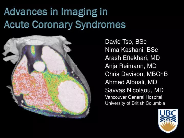
David Tso, BSc Nima Kashani, BSc Arash Eftekhari, MD Anja Reimann, MD Chris Davison, MBChB Ahmed Albuali, MD Savvas Nicolaou, MD Vancouver General Hospital University of British Columbia. Advances in Imaging in Acute Coronary Syndromes. Objectives.

E N D
David Tso, BSc Nima Kashani, BSc Arash Eftekhari, MD Anja Reimann, MD Chris Davison, MBChB Ahmed Albuali, MD Savvas Nicolaou, MD Vancouver General Hospital University of British Columbia Advances in Imaging in Acute Coronary Syndromes
Objectives • To review the imaging modalities available in assessing patients with acute coronary syndrome (ACS) • To summarize the clinical trials investigating Multi-detector CT (MDCT) in diagnosing ACS • To discuss the benefits of MDCT in assessing ACS with regards to cost, time to diagnosis, outcomes • To discuss the role of a Triple-Rule-Out Protocol in evaluation of chest pain syndromes
Cause for concern • ACS is associated with increase in cardiac death and subsequent MI • 2-8% of patients with ACS are misdiagnosed and inappropriately discharged home, which is associated with doubling mortality rate • Important to differentiate serious causes of chest pain from less serious causes • Angina • Pulmonary embolism • Aortic dissection Christenson J, et al. CMAJ 2004 Jun 8;170(12):1803-7 Chinnaiyan KM, Raff GL, Goldstein JA. Cardiol Clin. 2009 Nov;27(4):587-96. White CS, Kuo D. Radiology. 2007 Dec;245(3):672-81.
Definition of ACS • Constellation of clinical symptoms that are compatible with acute myocardial ischemia • STEMI & NSTEMI • Unstable Angina (UA) • UA/NSTEMI • ECG ST-segment depression or prominent T-wave inversion • +/- Positive biomarkers of myocardial necrosis J Am Coll Cardiol. 2007 Aug 14;50(7)
Standard of Care • Clear evidence of STEMI • Suggestive clinical history & exam • ST-elevation on ECG • Positive Cardiac biomarkers • Consider immediate reperfusion therapy • Fibrinolysis • Percuntaneous coronary intervention • Extremely low probability of ACS • Discharge J Am Coll Cardiol. 2007 Aug 14;50(7)
Management based on work-up • Chest pain indeterminate at initial work-up • Atypical clinical history & exam • ECG showing only non-specific T-wave changes • Normal biomarkers • Further diagnostic evaluation required • Rest myocardial perfusion imaging w/ SPECT • Stress echocardiography • MRI • MDCT White CS, Kuo D. Radiology. 2007 Dec;245(3):672-81. J Am Coll Cardiol. 2007 Aug 14;50(7)
SPECT Benefits vs. Limitations BENEFITS • Highly sensitive • 90-100% • Moderate specificity • 60-78% • High negative predictive value • 97-100% • Good prognostic value LIMITATIONS • High radiation exposure • Nuclear medicine near ED • Only assesses CAD and not other causes • Time intensive • Potential for false negatives • Provides no anatomical information Reza Fazel et al. N Engl J Med, 27 Aug 2009, 361(9):849. White CS, Kuo D. Radiology. 2007 Dec;245(3):672-81.
Echo: Benefits vs. Limitations BENEFITS • No radiation exposure • Similar sensitivity and specificity as radionuclide perfusion imaging • Portability LIMITATIONS • Off hours availability • False-negative results in patients with small myocardial infarctions or unstable angina • May fail to identify non-structural infarcts • Might have ischemia but no wall abnormality • Limited anatomical information White CS, Kuo D. Radiology. 2007 Dec;245(3):672-81.
Cardiovascular Magnetic Resonance • Accepted indications for assessment by CMR • Congenital heart disease • Great vessels • Acquired myocardial & pericardial disease • CAD • Role in ACS less well established • CMR may be useful in the acute setting as a problem solving tool • Patients with suspect ACS but no angiographic evidence of coronary artery stenosis • Utility in negative or equivocal findings on CT • Establish degree of myocardial necrosis after establishing MI Scirica BM. J Am Coll Cardiol. 2010 Apr 6;55(14):1403-15. Lockie et al, Circulation. 2009;119:1671-1681
MDCT in the Acute Care Setting • Provides excellent spatial resolution provides superior information of anatomy • Provides functional information through blood perfused volume and stress protocols • Ability for plaque analysis • Appropriate use of Triple-Rule-Out Protocol can explore other differential diagnoses for chest pain • MDCT imaging protocols incorporated into ACS workup demonstrates savings in time to diagnosis, costs while providing good patient outcomes
CT Angiography • Direct visualization of coronary arteries was previously limited to invasive techniques • I.e. coronary angiography • Introduction of Multi-detector CT (MDCT) in non-invasive evaluation of CAD has become possible • MDCT performs well in detection of significant coronary stenosis • Sensitivity = 82-95% • Specificity = 82-98% • Presence of coronary calcifications in patients with ACS shown to be predictive of future cardiac events J Am Coll Radiol. 2006 Oct;3(10):751-71.
Benefits of MDCT • Performs well in ruling out CAD for low to intermediate probability of CAD • High negative predictive values • Patients with normal scan may be discharged safely • CCTA may not provide additional relevant diagnostic information in patients with a high pretest probability for CAD • May need further investigations because of low positive predictive value • Test of choice = Conventional coronary angiography
MDCT Limitations • Image quality suffers from fast heart rate • Requires premedication with β-blockers • Arrhythmias, ectopy, or ECG artifacts result in degradation of image quality • ECG-gating critical to coronary imaging • Radiation dose to patient • Provides anatomic information, but not physiologic data Chinnaiyan KM, et al. Cardiol Clin. 2009 Nov;27(4):587-96.
MDCT: Cost & time to diagnosis Using CCTA vs. standard of care protocols (i.e. myocardial perfusion imaging) can diagnosis patients faster Cost savings come from reduce time in hospital and reduced need for additional tests from a negative CCTA exam Goldstein et al. J Am Coll Cardiol 2007;49:863–71 May et al. AJR 2009; 193:150–154
Ruling out Non-cardiac Causes • Routine CT acquisition has ability to examine other non-cardiac structures • e.g. Aorta, pulmonary arteries • Possible modality to rule out potentially fatal causes of chest pain • CAD • Acute aortic dissection • Pulmonary embolism • Triple rule out (TRO) protocol can allow in rapid discharge of patients with low to moderate ACS risk Chinnaiyan KM, et al. Cardiol Clin. 2009 Nov;27(4):587-96.
Triple rule out: atypical chest pain Diagnosis = pulmonary embolism
TRO – Pericardial Effusion • 40 yo female • Atypical chest pain • SOB Diagnosis = Lymphoma resulting in pericardial effusion
Whole Body Rule-out Diagnosis = Aortic Dissection
Dedicated CTA vs. Triple-Rule-Out • Investigates coronary arteries only • Greater spatial resolution of coronary arteries • Less radiation • Less contrast • Time = 8 secs • Craniocaudal • Investigates CAD, PE, Aortic dissection • Lesser spatial resolution of coronary arteries • More radiation • More contrast • Time = 15 secs • Caudalcranial Dedicated CTA Triple-Rule-Out
Heart perfused volume imaging • Myocardial blood pool analyzed by assessing iodine content within myocardium • Using unique X-ray absorption characteristics of iodine at different kV levels • Color-coded “iodine maps” represent myocardial blood pool • Perfused myocardium contains iodine vs. an infarct which will not have iodine uptake • Single cardiac CTA exam that examines both coronary anatomy and myocardial perfusion is promising Rocha-Filho et al. Radiology: Volume 254: Number 2—February 2010 Ruzsics et al. Eur Radiol. 2008 Nov;18(11):2414-24.
Dual energy CT + adenosine stress • Recent studies show results from adenosine-mediated CT perfusion imaging is comparable to SPECT–myocardial perfusion in detecting perfusion abnormalities • Allow for comparison of rest and stress DECT in detecting perfusion deficits • Protocol allows for quantification of iodine • DECT adenosine stress protocol enables examination of anatomy and function in a single investigation • Radiation exposure equivalent to SPECT • Regadenoson = selective α2a receptor agonist • Coronary vasodilator • Less side effects than adenosine • Easier to use iv bolus 5cc(0.4 mg) with no weight adjustment
CTA + CT Heart Perfused Blood Volume • Combination of cardiac CT angiography and CT perfusion in a single examination improved diagnostic accuracy • Comparable to SPECT–MPI • For stenosis > 50% luminal narrowing • Combination shown to increase PPV by more than 20% after incorporation of CT perfusion analysis over CTA alone (66% to 86%) • Myocardial hypoenhancement seen on MDCT has potential in evaluating CAD without additional cost in radiation dose or contrast load. Kachenoura N, et al. Am J Cardiol. 2009 Jun 1;103(11):1487-94. Rocha-Filho et al. Radiology: Volume 254: Number 2—February 2010
Perfusion defects at rest anterior LAD lateral LCx posterior RCA 100% iodine overlay
100 kV (Stress) 50:50 heart perfused volume (Stress)
Heart perfused blood volume at rest 50:50 100% iodine overlay
DE – Heart blood perfused volume Stress Perfusion Rest Perfusion
Acute Chest Pain Algorithm MDCT = One Stop Shop Triple Rule-Out Chinnaiyan KM, et al. Cardiol Clin. 2009 Nov;27(4):587-96.
Conclusions • MDCT has a role as a multipurpose triaging tool in assessing patients with atypical chest pain • MDCT has been proven in clinical trials to have great accuracy in ruling out ACS • MDCT in combination with stress perfusion may yield better diagnostic accuracy • Appropriate use of Triple-Rule-Out Protocol can explore other differential diagnoses for chest pain • MDCT imaging protocols incorporated into ACS workup demonstrates savings in time to diagnosis, costs while providing good patient outcomes
CTA scans use Test Bolus of 6.5cc/sec for 65 cc isovue 370, followed by a 60/40 split bolus of saline/isovue 370, followed by 40cc of pure saline. Peak HU for contrast is determined at ascending aorta, and 5-6 sec is added for delay time for scan after contrast flow is started at the R.ACF