Download
1 / 25
430 likes | 1.33k Views
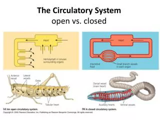
The Circulatory System open vs. closed. Red = oxygen-rich Blue = oxygen-poor. Cardiovascular system components: heart 2 atria, 2 ventricles blood vessels arteries, arterioles, capillaries, venules , veins blood Pulmonary circulation— to gas exchange tissues (lungs)

E N D
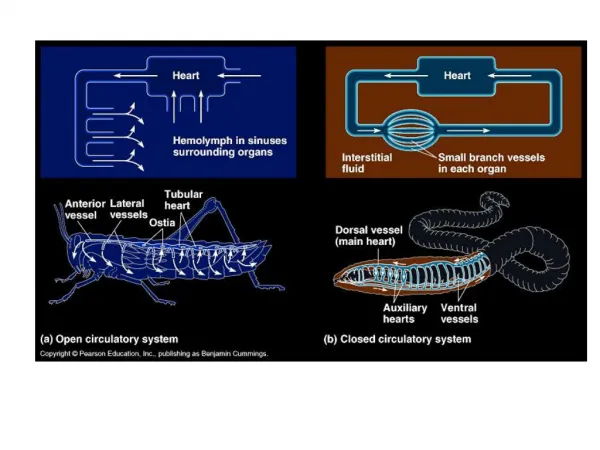
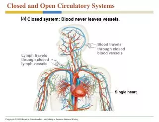
The Circulatory System open vs. closed
Cardiovascular system components: • heart • 2 atria, 2 ventricles • blood vessels • arteries, arterioles, capillaries, venules, veins • blood • Pulmonary circulation— to gas exchange tissues (lungs) • Systemic circulation— to body tissues • Double circulation allows blood to be pumped with higher pressure to organs.
Pulmonary artery has deox. blood! Pulmonary vein has ox. blood! Right ventricle—goes to pulmonary circuit Left ventricle—goes to systemic circuit
4 valves prevent backflow of blood in the Atrioventricular (AV) valves are between the atria and the ventricles—keep blood from going from ventricles BACK to atria Semilunar valves are between the left ventricle and the aorta and between the right ventricle and the pulmonary artery—keep blood from going from arteries BACK to ventricles Heart murmur = defect in a valve
“Lub” = AV valves close (ventricles contract—pumping) This is called systole. “Dup” = semilunar valves close (ventricles relax—filling) This is called diastole The cardiac cycle is one systole and one diastole.
The sinoatrial (SA) node (aka pacemaker) sets the rate at which cardiac muscle cells contract. It’s located in the wall of the right atrium. It generates electrical impulses, and because the cardiac muscle cells are all connected, they contract in unison. The AV node delays the impulse so that the atria contract before the ventricles.
Nerves, hormones, and temperature control the SA node.
Smooth muscle in the artery walls pumps blood. Veins rely on skeletal muscle around them to pump blood back to the heart. Veins have valves to prevent backflow.
Blood pressure is high in the arteries and low in the veins. BP is the hydrostatic force that blood exerts against the wall of a vessel.
RBCs have no nuclei, look like life savers, and have no mitochondria. So they can only make ATP by anaerobic means! This is so they don’t use up the oxygen they carry…pretty clever! RBCs have lots of hemoglobin—hemoglobin picks up oxygen in the lungs and carries it to all other organs.
Pluripotent stem cells can differentiate into any type of blood cell. These cells are found in red bone marrow. Erythropoietin, made in the kidney, stimulates production of RBCs.
Gas Exchange—Respiration Uptake of oxygen from the environment Discharge of carbon dioxide to the environment Remember that oxygen is used for cell respiration and so is constantly being used up. And carbon dioxide is the waste product of cell respiration.
Respiratory surface is where the environmentmeets the cells—cellsmust be bathed in water, so the surfaces are moist.
Countercurrent exchange in fish gills: Water flowsoppositeto blood flow. Countercurrent exchange allows blood to pick up more oxygen than concurrent flow.
Our respiratory surfaces (alveoli) are folded inside our bodies so that we don’t lose water. We have lungs! (as do reptiles and birds)
Breathing enables us to maintain a high oxygen concentration and a low carbon dioxide concentration. We breathe using negative pressure—like a suction pump pulling air into the lungs, which are enclosed by a double-walled sac. lungs diaphragm
Breathing control centers are located in the pons and the medulla of the brain. Sensors in the medulla and in the carotid arteries in the neck detect changes in CO2 concentration (blood pH) to direct control of breathing ↑CO2 ↓blood pH
O2 is collected from the lungs by hemoglobin in blood and is dropped off in body tissues. CO2is collected from tissues and is deposited in the lungs to be expelled.
Hemoglobin is more likely to release O2 to tissues that don’t have enough. The Bohr Shift—More O2 is released from hemoglobin when there’s more CO2 (lower pH)
Most CO2is transported in blood in the form of HCO3- (bicarbonate ions). Conversion of bicarbonate ions to carbonic acid and vice versa maintains the pH of blood To lungs →