Download
1 / 36
360 likes | 523 Views
Initial Resting Assessments. Blood Pressure, Heart Rate, Cholesterol. Resting ECG. Used to determine if arrythmias are present Used to assess HR General assessment. Electrocardiogram (ECG). w Records the heart's electrical activity and monitors cardiac changes.

E N D
Initial Resting Assessments Blood Pressure, Heart Rate, Cholesterol
Resting ECG • Used to determine if arrythmias are present • Used to assess HR • General assessment
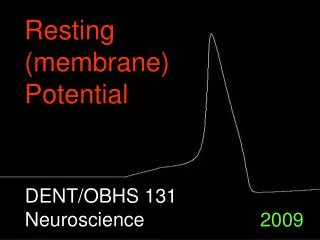
Electrocardiogram (ECG) w Records the heart's electrical activity and monitors cardiac changes w The P wave—atrial depolarization w The QRS complex—ventricular depolarization and atrial repolarization w The T wave—ventricular repolarization
Assessing Blood Pressure Classifications for Exercise
BP • The three most important variables affecting BP • 1. Stroke Volume • 2. Total Peripheral Resistance • 3. Heart Rate • BP= HR x SV x TPR
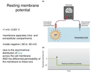
BP • A measure of the pressure exerted by the blood on the arteries • systolic BP - the pressure in the arteries during systole (the contractile phase of the cardiac cycle) • diastolic BP - the pressure in the arteries during diastole (the relaxation phase of the cardiac cycle)
Cardiac Cycle w Events that occur between two consecutive heartbeats (systole to systole) w Diastole—relaxation phase during which the chambers fill with blood (T wave to QRS) w Systole—contraction phase during which the chambers expel blood (QRS to T wave)
Cardiac Cycle • A. Systole • 1. Isovolumetric (no change in volume) Contraction • ventricles are excited and begin to contract (no blood moving) • valves are closed • ventricular P is
Cardiac Cycle • Systole (cont.) • 2. Ejection • when P in vent>aorta (~120mmHg) • semilunar valves open • blood ejected from ventricles into aorta and pulmonary artery
Cardiac Cycle • B. Diastole • 1. Isovolumetric Relaxation • ventricles are relaxed • semilunar valves closed • ventricular P is • atrial P<vent P • vent P<aorta P
Cardiac Cycle • Diastole (cont.) • 2. Filling • AV valves open • blood flows into vent • vent P<atrial P
BP** • Resting Systolic 110-140 mmHg • Resting Diastolic 60-90 mmHg
BP Assessment (ACSM) Blood Pressure: Systolic Diastolic Optimal <120 <80 Normal 120-129 80-84 High Normal 130-139 85-89 Hypertension: Stage 1 140-159 90-99 Stage 2 160-179 100-109 Stage 3 >180 >110
Methods of Assessing BP • Auscultation using a stethoscope and sphygmomanometer • 1. Seated for at least 5 minutes with arm the level of the heart (no caffeine or smoking 30 min prior) • 2. Align cuff with brachial artery (the bladder should encircle 80% of an adults arm and 100% of a child’s arm)
Methods of Assessing BP • 3. Place stethoscope bell over the brachial artery beneath the cuff • 4. Inflate cuff quickly to 20 mmHg above estimated systolic • 5. Slowly release valve (2-3mmHg/s) noting first Korotkoff sound)
Methods of Assessing BP • 6. Continue releasing until sound becomes muffled (4th) and then disappears (5th) • 7. Wait 30s and repeat (use the average)
BP Sounds • The sound of blood moving through the vessels is normally silent. • Smooth laminar blood flow - blood in center of vessels moves faster than blood closest to vessel walls (produces little sound)
BP Sounds • Pinching the artery causes turbulence and is noisy • The tendency of the cuff pressure to constrict the artery is opposed by blood pressure • If cuff pressure is greater than systolic pressure the artery is completely constricted and no sounds are heard.
BP Sounds • When pressure is released from the cuff the first sound you hear (1st Kortokoff sound) is when the cuff pressure reaches the systolic pressure • Blood is passing turbulently as the artery becomes unconstricted
BP Sounds • You continue to hear sounds at every systole (contraction of the heart) as long as the cuff pressure remains above diastolic pressure • When sound becomes muffled is called the 4th Kortokoff sound (7-10 mmHg higher than 5th Kortokoff sound)
BP Sounds • When cuff pressure reaches diastolic pressure the sounds disappear (5th Kortokoff sound) since the artery opens and laminar blood flow begins • Use 5th sound as an index of diastolic pressure
Heart Rate Assessment Classifications for Exercise
HR Assessment • Methods • Auscultation - using stethoscope, count beats for 30-60 seconds • Palpation - at brachial, carotid, radial, or temporal artery • Heart Rate Monitors • ECG
Palpation • Use tips of index and middle fingers (not the thumb) • Don’t apply heavy pressure to carotid (baroreceptors will slow the heart) • Count beats for 6 (x10), 10 (x6), 15 (x4), 30 (x2), or 60 second (6 and 10 when exercising or immediately post-exercise; 15, 30, 60 for resting)
Assessing Cholesterol Classifications for Exercise
Fats/Cholesterol • Fats = triglycerides and cholesterol • Cholesterol = fat-like substance found in foods of animal origin • Triglycerides = three fatty acids attached to a glycerol molecule • saturated or unsaturated
Lipoproteins • Transport fat-like substances (cholesterol/triglycerides) through the blood • Very Low Density Lipoproteins (VLDL) - major carrier of triglycerides • Low Density Lipoproteins (LDL) - major carrier of cholesterol deposited on artery walls.
Lipoproteins • High Density Lipoproteins (HDL) - carry cholesterol to liver to be disposed of
Cholesterol Measures • 1. Total Cholesterol = VLDL + LDL + HDL • Desirable < 200 mg/dl • Borderline high 200-239 • High >240
Measures • LDL • Desirable <130 • Borderline 130-159 • High >160
Measures • Triglycerides • Desirable <200 • Borderline 200-400 • High 400-1000 • Very High >1000
Measures • HDL • Low <35 • Normal 35-60 • High >60
Measures • TC/HDL Ratio • ideal <3.5 • risk 3.5-5 • higher risk - greater than 5 • Page 19 (Heyward) • Pae 44-45 (ACSM)
Blood Profile • Blood Glucose • Hemoglobin • Hematocrit • Potassium • Blood Urea Nitrogen • Creatinine • Iron • Calcium