Download
1 / 24
430 likes | 883 Views
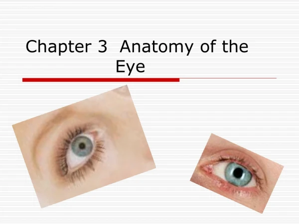
Anatomy of the Eye. Introduction. The eye is a specialized sense organ that helps us to understand our environment. It is a sensory unit composed of three parts: receptor, sensory pathway, and a brain center. . Introduction. In the eye, the following occurs:

E N D
Introduction The eye is a specialized sense organ that helps us to understand our environment. It is a sensory unit composed of three parts: receptor, sensory pathway, and a brain center.
Introduction • In the eye, the following occurs: -photoreceptors in the eye convert light energy into nerve impulses -nerve impulses are transmitted along the optic nerve -the impulses are interpreted as vision in the occipital lobe of the cerebrum
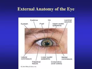
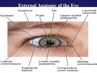
Accessory Organs & Eye Protection • Orbital cavities (bony sockets) – house & protect the eye • Adipose tissue – cushions the eye and absorbs shock from blows
Accessory Organs & Eye Protection • Lacrimal glands – produce tears that lubricate & have a germicidal effect • Eyebrows – protect against foreign articles, perspiration, & direct rays of light
Accessory Organs & Eye Protection • Eyelids – folds of skin that cover the surface of the eye; close by reflex action when an object approaches • Eyelashes – secrete oils that prevent lids from sticking together
Accessory Organs & Eye Protection • Extrinsic muscles – muscles located outside of the eye that control certain eye movements such as moving the eyeball from side to side or rolling the eyes
Accessory Organs & Eye Protection • Intrinsic muscles – muscles located inside the eye that help hold the lens in place & modify its shape
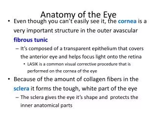
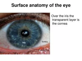
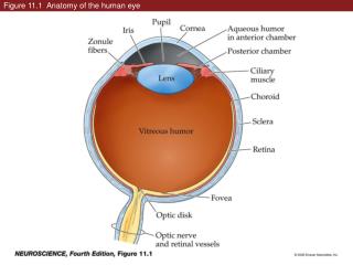
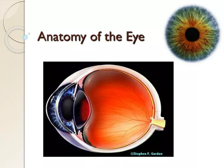
Eye Parts • Sclera – white, outer layer of the eyeball; tough, fibrous membrane that helps to maintain the spherical shape of the eyeball • Cornea – part of sclerotic coat; transparent, front part of eyeball through which light waves pass – no blood vessels but lots of nerve endings
Eye Parts • Canals of Schlem – venous passages that drain the fluid that accumulates behind the cornea; located where the sclera & cornea meet • Conjuctiva – thin, transparent mucous membrane that covers the eyeball
Eye Parts • Choroid layer – middle layer of the eye; supplies blood vessels to the eye and contains dark pigment granules that prevent the reflection of light in the eye
Eye Parts • Ciliary body – intrinsic muscle; smooth muscle fibers support & modify lens shape • Iris – colored portion of eye formed by circularly and radially arranged smooth muscle fibers; regulates amount of light entering they eye by constricting or dilating the pupil
Eye Parts • Pupil – rounded opening of the iris through which light passes • Retina – innermost layer of the eye; lines its surface and contains photoreceptors (cells responsible for converting light into nerve impulses – rods & cones)
Eye Parts • Rods – cylindrical photoreceptors found in greatest concentration on the edges of the retina; most common type of receptor; allow us to see in low light and provide for peripheral vision
Eye Parts • Cones – Conical photoreceptors found in greatest concentration near the center of the retina; there are three varieties of cones, each most sensitive to a particular wavelength (color) of light – blue, green, & red; allow for visual acuity (sharp vision) and color vision
Eye Parts • Fovea centralis – a depression, or pit, in the center of the retina that contains only cones; provides for the most acute vision & color sensitivity • Optic disk (blind spot) – area where optic nerve attaches to the retina; does not contain any photorecptors
Eye Parts • Lens – flexible, biconvex, crystal-like structure that brings rays of light into focus and produces an image on the retina
Eye Parts • Suspensory ligament – holds the lens in place; attached to the ciliary body, which controls the amount of tension exerted on the lens
Eye Parts • Aqueous humor – watery fluid that provides nutrition and helps maintain the shape of the cornea; found in the smaller, anterior chamber of the eye
Eye Parts • Vitreous humor – thick, gel-like substance that fills the largest chamber of the eye and helps to hold its spherical shape
Tapetum Iridescent layer in eye that causes glow in dark appearance when light shines in - many animals have this