Download
1 / 10
100 likes | 283 Views
Your Lungs. A Complete Guide. ^. Almost. Your Lungs. The Respiratory System.

E N D
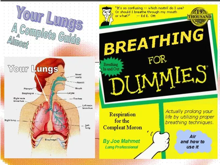
Your Lungs A Complete Guide ^ Almost Your Lungs
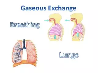
The Respiratory System The primary function of the respiratory system is to supply the blood with oxygen, in order for the blood to deliver oxygen to all parts of the body to carry out respiration. The respiratory system does this through breathing. When we breathe, we inhale the air from our environment notably the most important oxygen though this only accounts for around 21% of the earths atmosphere. and exhale waste gases from our bodies most importantly Carbon Dioxide but there are others such as carbon monoxide and others. This exchange of gases is the respiratory system's means of getting oxygen to the blood, and therefore all round the body.
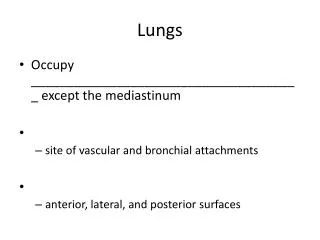
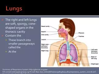
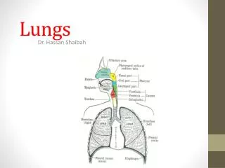
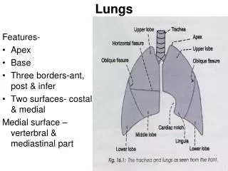
Organs in the Respiratory System Trachea –(also know as wind pipe) is where air is funnelled down from the mouth towards the lungs, also mucus is transported up here to be swallowed. Bronchi –(left and right) are the entrances to your lungs these separate from the trachea into your left and right lung Bronchioles –separating out from the Bronchus these are considerably smaller and become smaller and smaller until you reach a single alveoli Alveoli –these are on the very end of the bronchioles and are like balloons (inflatable with a very thin wall/membrane. Pleura –(pleurae) are the membranes connecting the lungs to the chest wall, the inner one is attached to the lungs, the outer to the chest wall and inside is a gap or space known as the pleural cavity Diaphragm –this is a layer of muscle that moves up and down to make the lungs inhale and exhale air. Hiccupping (Hiccough). Yes your diaphragm is responsible for you hiccupping when you hiccup your diaphragm involuntarily spasms causing a sudden rush of air into your lungs force your epiglottis to close creating the “hic” sound. Question What else apart from breathing is the Diaphragm responsible for?
The Trachea The trachea, or windpipe, is a tube that has an inner diameter of about 20-25 mm and a length of about 10-16cm. It extends from the Larynx to the primary (main) bronchi in Mammals allowing the passage of air to the lungs. In humans there are about 15 – 20 incomplete C-shaped cartilaginous rings which reinforces the anterior and lateral sides of the trachea to protect and maintain the airway open. There is a piece of smooth muscle connecting the ends off the incomplete cartilaginous rings called the Trachealis muscle. This contracts reducing the size of the lumen of the trachea to increase the air flow rate during coughing
Question The Left Lung is Divided into two lobes, but how many is the right divided into? Three, yes surprisingly while the left lung is divided into two lobes the right is divided into three. Bronci (Broncus) and Bronchioles Where the Trachea splits into the bronchus (left and Right) and (left) a primary Bronchi splitting into a secondary bronchi. The trachea (windpipe) divides into two main bronchi the left and the right, at the level of the sternal angle. The right main bronchus is wider, shorter, and more vertical than the left main bronchus. There is hyaline cartilage present in the bronchi, present as irregular rings in the larger bronchi (and not as regular as in the trachea), and as small plates and islands in the smaller bronchi. Smooth muscle is present continuously around the bronchi. Cross section of a bronchiole
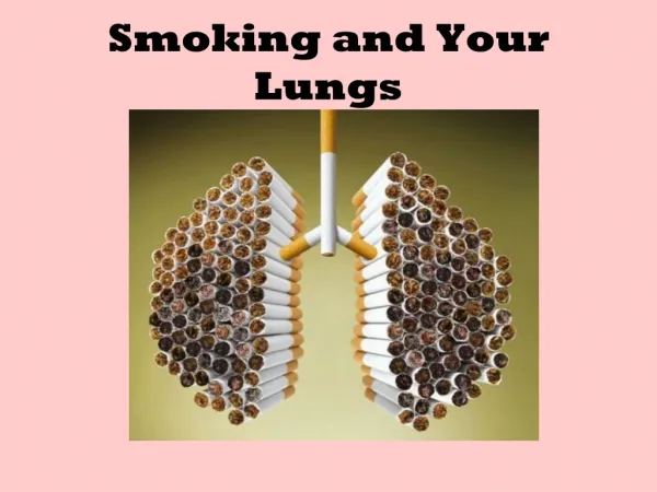
Cells in The Trachea, Bronchi and Bronchioles The trachea is line with lots of tightly packed cells with cilia on the top part facing the inside of the trachea. Below the cells the submucus layer contain mucus glands these produce mucus which rises up through the cells to line the trachea to catch the bacteria and things we don’t want in out lungs when we breathe in, as they come in with the oxygen.They have cilia move the mucus around the lining of lungs up to the Larynx where it will be swallowed and digested. The cells are positioned on tops of layers of other things such as , Cilia • Elastic fibres • Submucus layer • Fibrous membrane The cell (left) has been dyed so different parts are easier to see
Question Does The Pulmonary Artery carry oxygenated or deoxygenated blood? Blood Vessels in the Lung This diagram shows the main route the “Pulmonary Artery” and the “Pulmonary Vein” takes. It has an almost piercing effect through the lung It is a major part of oxygen uptake. And in supplying the lungs with blood. The pulmonary artery and pulmonary vein are the major source of getting, and taking away blood from the lungs. Like the heart it has two large blood vessel the artery and vein. However unlike the heart they are the other way around the artery carries deoxygenated blood (they are the only arteries apart from umbilical arteries in foetuses‘ that carry deoxygenated blood) and the vein carries oxygenated blood back to the heart. (this is the same as the artery no other vein does this except foetal veins)
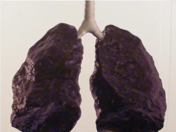
Alveoli and Alveolar Ducts The walls of Alveoli are composed of only a single layer of cells, the endothelium. This layer is so thin that molecules such as oxygen and water can pass through them by diffusion and enter the Capillaries which also only have a layer around them one cell thick. Waste products such as carbon dioxide and Carbon Monoxide can diffuse back into the Alveoli to be carried away for removal from the body. Capillaries are also very narrow (small) and they are so small the blood cells need to pass through it them in a single file line.
CO2 O2 Blood Capillary Gas Exchange (Respiration) Respiration this name can cause problems - in biology the word "respiration" can mean cellular respiration or metabolism (ATP generation inside cells), however sometimes (such as here) it can also refer to breathing (which is how the word is most often used by non-biologists). • Gases cross the respiratory surface by diffusion, so from Fick's law we can predict that respiratory surfaces must have: • a large surface area • a thin permeable surface • a moist exchange surface • Alveoli have all of these they are specifically made to increase diffusion through the alveolar walls. Capillary
Facts You May Not Know... Question 1500 miles, that’s enough to reach from the very south of Britain to the very north, and back. How Many Miles of airways do you lungs contain? Question 300 – 500 million, you have around 300 – 500 million alveoli in your lungs How Many Alveoli do your lungs contain? Question 75m2 that’s about the area of a tennis court. And what area do those alveoli cover? Question 620 miles, that’s the length of Britain from edge to edge. If the capillaries in your lungs were unwound how many miles would they cover?